TL;DR: A 2026 mouse and cell study in Nature found that developing neurons can generate DNA double-strand breaks while moving through narrow brain tissue during migration, then usually repair those breaks without cell death.
Key Findings
- Developing neurons: The study tracked cerebellar granule neurons, Purkinje cells, and cortical neurons during normal brain development.
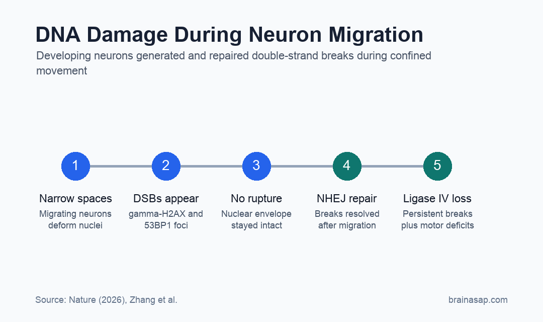
- Confined migration: Neurons formed DNA damage markers while moving through narrow spaces and microfabricated constrictions.
- No nuclear rupture: Unlike many cancer cells, cerebellar granule neurons generated breaks without detectable nuclear-envelope rupture.
- TOP2beta result: Confined migration increased topoisomerase-IIbeta-bound double-strand breaks.
- Ligase IV risk: Removing a key repair enzyme caused persistent DNA damage, transcriptional changes, and mild later-life motor deficits in mice.
Source: Nature (2026) | Zhang et al.
Neuron migration is a normal part of brain development. Newborn neurons have to move from where they are generated to where they will become part of mature circuits.
When developing neurons compress their nuclei while moving through tight spaces, they can form DNA double-strand breaks, or DSBs, even during normal development.
Migrating Neurons Generated DNA Break Markers During Brain Development
The researchers focused first on cerebellar granule neurons, which migrate during the first postnatal weeks in mice. These neurons move from the external granule layer through the molecular layer to the internal granule layer.
Using gamma-H2AX and 53BP1, two common markers of DNA damage response, the team observed DSB signals in postmitotic neurons during migration. The signals appeared in the molecular layer and internal granule layer, where migrating neurons were moving through tissue.
- Timing: DNA damage markers in postmitotic cerebellar granule neurons were visible from postnatal day 4 through postnatal day 15.
- Resolution: By postnatal day 30, gamma-H2AX foci were no longer detectable in the same developmental context.
- Cell types: Similar damage markers appeared in migrating Purkinje cells and cortical neurons.
- Live imaging: 53BP1 foci formed transiently as neurons migrated in organotypic slice cultures and constricted channels.
Migrating neurons were not dying in large numbers in these assays. Instead, migration created DNA lesions that a healthy developing brain normally has to repair.
Microchannel Experiments Linked Tight Spaces to DNA Damage
To test whether mechanical confinement was enough to trigger the damage response, researchers placed neurons in microfabricated channels of different widths. Narrower channels increased nuclear deformation during movement.
In 3-micrometer channels, cerebellar granule neurons formed 53BP1 foci during and after passage through the constriction. The foci were transient, which is consistent with damage detection followed by repair.
Transwell experiments gave a second test. More than 20% of neurons carried multiple gamma-H2AX or 53BP1 foci at 6 and 12 hours after migration through 3-micrometer pores, but levels returned toward control by 24 hours and later.
- Confined pores: 3-micrometer migration produced more DNA damage markers than larger 8-micrometer pores.
- Postmitotic cells: The result persisted after researchers depleted dividing precursor cells, supporting a migration-related mechanism.
- Low apoptosis: TUNEL assays showed little to no increase in apoptotic cell death up to 36 hours.
Confined migration created detectable DNA damage, but the cells usually survived and resolved the damage response.
Neurons Formed Breaks Without Nuclear-Envelope Rupture
In many cancer or immune-cell models, tight migration can rupture the nuclear envelope, letting cytoplasmic factors enter the nucleus and increasing DNA damage. The neuronal result was different.
Using nuclear-localization reporters and cGAS markers, researchers saw nuclear-envelope rupture in HeLa cells under confinement. In cerebellar granule neurons, rupture was not detected even when nuclei were severely deformed in 2- or 3-micrometer channels.
- HeLa comparison: Confined HeLa cells showed reporter leakage consistent with nuclear-envelope rupture.
- Neuron comparison: Migrating cerebellar granule neurons kept nuclear-envelope integrity under similar constriction.
- Rare rupture events: Nuclear blebbing or rupture occurred in less than 3% of granule neurons migrating through 3-micrometer pores.
The mechanism therefore appears different from the better-known rupture model. Developing neurons can generate DSBs during confinement without tearing open the nuclear envelope.
TOP2beta and Non-Homologous End Joining Shaped the Repair Path
The researchers next tested topoisomerase IIbeta, or TOP2beta, an enzyme that helps manage DNA torsional stress. Confined migration increased TOP2-DNA cleavage complexes, consistent with enzyme-bound breaks.
Repair also appeared to use non-homologous end joining, a DSB repair pathway often shortened as NHEJ. The repair marker pattern included Ku70 near gamma-H2AX nanodomains, while RAD51 foci, a homologous recombination marker, were not observed in cerebellar granule neurons.
The repair pathway became consequential when the team removed ligase IV, a key NHEJ enzyme, at the onset of neuronal migration. Persistent DSBs accumulated in cerebellar neurons.
Those mutant mice later showed moderate transcriptional changes in genes related to synaptic function, neuronal development, stress responses, and immune responses. They also developed mild motor deficits later in life.
Normal Development May Include Repairable Genome Stress
The study reframes neuronal migration as both a placement process and a genome-maintenance challenge. Mechanical movement through dense tissue can create DNA damage, and normal development depends on repair systems clearing it.
The strongest interpretation is specific to the model. These experiments were done in mice, cultured neurons, microchannels, and genetic repair perturbations, not in human infants or patients with neurodevelopmental disease.
The disease relevance is still plausible. Neurons have limited regenerative capacity, and unresolved DNA damage appears in brain aging and neurodegeneration research.
The study adds a developmental route by which genome stress can arise before mature circuits are fully built. Migrating neurons can generate repairable DNA double-strand breaks during normal brain development, and faulty repair can leave measurable neural and motor consequences in mice.
Citation: DOI: 10.1038/s41586-026-10648-8. Zhang et al. Confined migration induces non-lethal DNA damage in developing neurons. Nature. 2026;654:1098-1108.
Study Design: Mouse brain-development study with organotypic slices, cultured neurons, microfabricated confinement assays, genome sequencing, and genetic repair disruption.
Sample/Model: Developing mouse cerebellar granule neurons, Purkinje cells, cortical neurons, and engineered migration assays.
Key Statistic: More than 20% of cerebellar granule neurons showed multiple gamma-H2AX and/or 53BP1 foci after confined 3-micrometer migration at 6 and 12 hours.
Caveat: The work is mechanistic and developmental; it does not show that the same repair failure occurs in a specific human disorder.