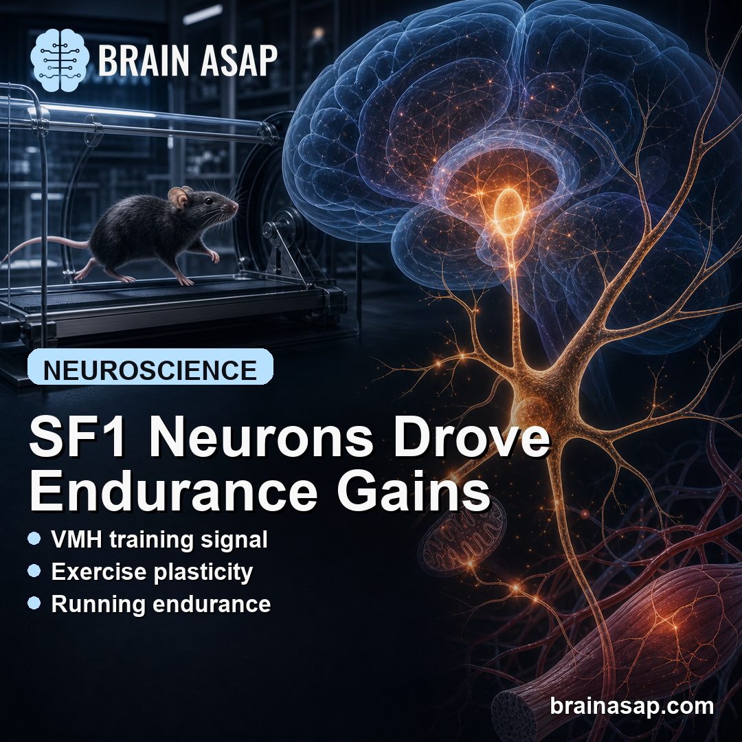
TL;DR: A 2026 mouse study in Neuron found that exercise activates ventromedial hypothalamic SF1 neurons, and that this brain response is required for normal endurance gains after training.
Key Findings
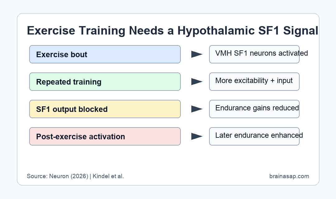
- VMH SF1 neurons: Exercise increased activity-linked Bdnf expression in steroidogenic factor-1 neurons in the ventromedial hypothalamus.
- Training plasticity: Repeated exercise increased both intrinsic excitability and excitatory synaptic input onto these SF1 neurons.
- Blocked output: Silencing VMH SF1 neuron transmission reduced endurance and running speed during exercise stress testing.
- Lost adaptation: Inhibiting SF1 neuron output after training blocked the usual endurance and metabolic gains from repeated exercise.
- Post-exercise stimulation: Activating SF1 neurons immediately after exercise enhanced later endurance, suggesting a central nervous system training signal.
Source: Neuron (2026) | Kindel et al.
Ventromedial hypothalamic SF1 neurons sit in a brain region that helps coordinate energy balance. The timing is the key detail: the circuit changed after exercise, when the body was preparing for later performance.
Exercise training is usually explained through muscle, heart, and metabolic tissue. In mice, researchers found a brain-side control point that was needed for normal performance gains and metabolic adaptation.
Exercise Activated SF1 Neurons in the Ventromedial Hypothalamus
The team focused on steroidogenic factor-1 neurons, usually shortened to SF1 neurons, in the ventromedial hypothalamus. These neurons are already known to respond to metabolic cues such as glucose, leptin, and insulin.
After a single exercise session, the researchers measured increased expression of Bdnf, an activity-linked gene, in VMH SF1 neurons. Repeated exercise then produced a stronger post-exercise activation pattern.
- Brain region: The ventromedial hypothalamus is a metabolic-control hub, not a muscle or cardiovascular tissue.
- Cell type: SF1-expressing neurons are excitatory neurons that help regulate energy use and whole-body metabolic state.
- Training history: Repeated exercise increased the number of activated SF1 neurons and the magnitude of activation after exercise.
This finding argues against a purely peripheral explanation for endurance training. The brain was not just receiving exercise information; it appeared to help organize the next round of adaptation.
SF1 Neuron Plasticity Increased After Repeated Exercise
Exercise training changed the electrophysiology of the VMH SF1 circuit. The researchers reported increased intrinsic excitability and a higher density of excitatory synaptic inputs onto SF1 neurons after repeated training.
Those changes suggest that exercise history was encoded as hypothalamic plasticity. In plain terms, the circuit became more responsive after repeated exercise exposure.
- Excitability shift: SF1 neurons were easier to activate after training.
- Synaptic input: The neurons received more excitatory drive, consistent with circuit remodeling.
- Timing: The relevant activation occurred after exercise, when the body is converting the bout into longer-term adaptation.
The study did not claim that this pathway is the only endurance mechanism. Skeletal muscle, blood vessels, metabolism, and endocrine systems still adapt to exercise.
The experiments indicate that those changes may require central coordination through the VMH SF1 circuit.
Silencing VMH SF1 Output Reduced Endurance Gains
To test causality, the researchers used genetic tools to disrupt VMH SF1 neuron transmission. Mice with blocked SF1 output had similar maximal oxygen consumption, but they showed reduced endurance and lower running speeds during an exercise stress test.
That distinction is important. The mice were not simply unable to consume oxygen. The deficit appeared in the coordinated performance and metabolic response that normally improves with training.
- Output blocked: Tetanus-toxin light chain was used to prevent synaptic vesicle release from VMH SF1 neurons.
- Performance effect: Endurance and running speed fell despite similar VO2 max.
- Training effect: Inhibiting SF1 output after training blocked the usual exercise-induced endurance improvement.
- Metabolic effect: The circuit also influenced energy-store use and skeletal-muscle remodeling markers during training.
The design makes the result stronger than a correlation. When SF1 signaling was disrupted, the endurance phenotype changed in the direction predicted by the activation data.
Post-Exercise SF1 Stimulation Enhanced Later Performance
The opposite experiment also mattered. When researchers stimulated SF1 neurons immediately after exercise, mice showed enhanced endurance performance later. That timing fits the idea that the post-exercise brain state helps initiate adaptation.
For human readers, the result should not be translated into a treatment claim. This was a mouse circuit study using invasive genetic and stimulation tools, not a human exercise trial.
- What the study supports: The central nervous system can be part of the biological machinery that converts exercise bouts into training gains.
- What it does not show: It does not identify a pill, supplement, or clinical stimulation protocol for endurance in people.
- Mechanistic meaning: Exercise adaptation may depend on brain-metabolism coordination, not only on local muscle remodeling.
The practical takeaway is mechanistic. A hypothalamic circuit that senses and responds to exercise may help coordinate how the body gets better at exercising again.
Mouse Circuit Tools Make the Mechanism Stronger but Narrower
The evidence is strongest because the researchers did not stop at measuring activation. They used SF1-Cre genetic tools, tetanus-toxin silencing, electrophysiology, and post-exercise stimulation to test whether the circuit changed endurance behavior.
Those tools also define the boundary of the result. The manipulations are precise in mice, but they are not equivalent to something a person can do during exercise training.
- Strong mechanistic evidence: Activation, plasticity, loss-of-function, and gain-of-function experiments pointed to the same VMH SF1 circuit.
- Model limitation: Mouse treadmill or stress-test endurance is not the same as human athletic performance, rehabilitation, or cardiometabolic health.
- Translation question: Future work would need to ask whether human hypothalamic activity markers track training adaptation in a measurable, noninvasive way.
Still, the study gives exercise biology a concrete brain-side target. It suggests that endurance training is partly a learned metabolic state coordinated by the central nervous system.
Citation: DOI: 10.1016/j.neuron.2025.12.033. Kindel et al. Exercise-induced activation of ventromedial hypothalamic steroidogenic factor-1 neurons mediates improvements in endurance. Neuron. 2026;114:1564-1575.e9.
Study Design: Mouse exercise-training and circuit-manipulation study.
Sample/Model: SF1-Cre mouse models with exercise stress testing, genetic silencing, electrophysiology, and neuronal stimulation.
Key Statistic: VMH SF1 neuron output was required for normal endurance gains, while post-exercise SF1 activation enhanced later endurance.
Caveat: The findings are preclinical and do not establish a human intervention.