TL;DR: A 2026 study in Nature Communications used cryo-electron microscopy and patch-clamp recordings to show how partially occupied GluK2/GluK5 kainate receptors can activate without entering the fast desensitized state that normally shuts glutamate receptors down.
Key Findings
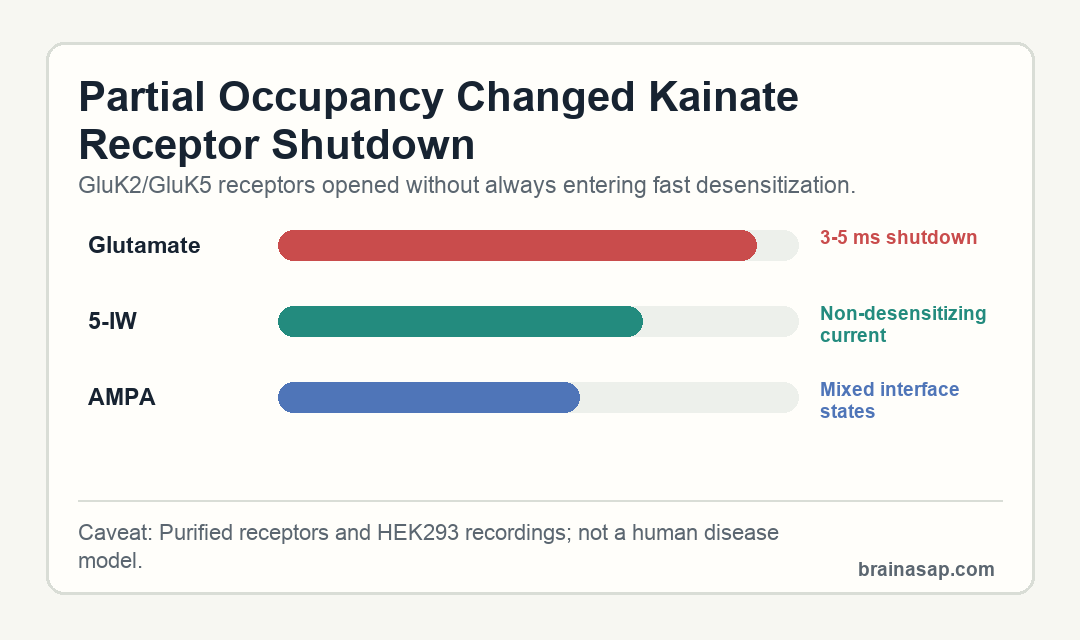
- Fast glutamate shutdown: Glutamate triggered receptor activation in less than 1 ms, followed by complete desensitization in about 3-5 ms.
- 5-IW behaved differently: The GluK5-selective agonist 5-iodowillardiine produced non-desensitizing currents in GluK2/GluK5 receptors but no measurable currents in GluK2 homotetramers.
- Two major cryo-EM classes: The 5-IW dataset separated into two similar particle classes, with 208,265 and 206,013 particles.
- Resolution reached receptor-domain detail: The 5-IW structures reached about 2.9-3.0 angstrom resolution for amino-terminal domains and 3.5-3.8 angstrom for ligand-binding/transmembrane modules.
- AMPA produced mixed states: AMPA-bound receptors showed partial desensitization and a range of intact, partially ruptured, and fully ruptured ligand-binding-domain interfaces.
Source: Nature Communications (2026) | Khanra et al.
Kainate receptors are ionotropic glutamate receptors, meaning they convert chemical glutamate signals into electrical current across neuronal membranes. This study focused on a heteromeric receptor made from GluK2 and GluK5 subunits, a combination that can respond differently from simpler receptor assemblies.
The central question was structural: how much agonist binding is enough to open the receptor, and how much is needed to push it into desensitization, the shutoff state that follows activation.
GluK2/GluK5 Kainate Receptors Separated Activation From Desensitization
Glutamate produced the expected sequence in HEK293 cells expressing GluK2 or GluK2/GluK5 receptors. Currents began in less than 1 ms, then rapidly desensitized in 3-5 ms.
The selective agonist 5-iodowillardiine (5-IW) gave a different result. It did not produce measurable current from GluK2-only receptors, but it produced substantial, non-desensitizing current from GluK2/GluK5 heterotetramers.
- Full glutamate response: Rapid activation was followed by complete fast desensitization.
- Partial GluK5-biased response: 5-IW activated GluK2/GluK5 receptors without triggering the same shutdown.
- Functional implication: Binding only part of the tetramer can be enough for current flow but not enough for the full desensitization rearrangement.
This distinction shapes synaptic timing because synaptic receptors do not simply turn on or off. Their occupancy state, subunit composition, and timing can change how long a synaptic signal lasts.
Kainate receptors are especially useful for this question because they can assemble from low-affinity GluK1-3 subunits and high-affinity GluK4-5 subunits. In a mixed GluK2/GluK5 receptor, selective binding at the GluK5 positions can probe only part of the tetramer instead of flooding every ligand-binding site at once.
5-IW Cryo-EM Structures Captured Two Similar Pre-Active States
Researchers purified GluK2/GluK5 receptors, exposed them to 5-IW, and froze samples within 1-2 minutes. The cryo-EM dataset separated into two major structural classes, with 208,265 particles in class 1 and 206,013 particles in class 2.
Both classes preserved the expected 2:2 stoichiometry: GluK5 occupied the A and C positions, while GluK2 occupied the B and D positions. The amino-terminal domains reached about 3.0 and 2.9 angstrom resolution, with lower but useful resolution in ligand-binding/transmembrane modules.
- Class 1: Amino-terminal domains were resolved at about 3.0 angstrom, and the LBD-TMD module at about 3.5 angstrom.
- Class 2: Amino-terminal domains were resolved at about 2.9 angstrom, and the LBD-TMD module at about 3.8 angstrom.
- Subunit assignment: Distinct glycosylation sites helped distinguish GluK2 from GluK5 inside the heterotetramer.
The main structural readout was the ligand-binding-domain interface. Instead of the fully ruptured arrangement associated with desensitization, the 5-IW condition preserved closed/open LBD dimers with intact interfaces.
AMPA Partially Desensitized GluK2/GluK5 Receptors
The AMPA experiments added a second partial-occupancy condition. AMPA is better known as an AMPA-receptor agonist, but in this system it acted as a GluK5-selective ligand and produced partial desensitization rather than the cleaner non-desensitizing 5-IW pattern.
Structurally, AMPA-bound receptors did not land in a single simple state. Researchers observed intact, partially ruptured, and fully ruptured ligand-binding-domain interfaces, which supports a stepwise view of receptor gating.
- Intact interfaces: Some receptors looked closer to a pre-active arrangement.
- Partially ruptured interfaces: Intermediate states suggested that the shutdown process can proceed unevenly across dimers.
- Fully ruptured interfaces: Other structures resembled the desensitized endpoint more closely.
This is the mechanistic center of the study. The same receptor family can encode different current timing depending on which subunits are occupied and how ligand-binding domains communicate across the tetramer.
The AMPA condition also helps avoid an overly simple interpretation. Partial occupancy was not automatically protective against desensitization; the ligand identity and the stability of specific LBD dimer interfaces both mattered.
Occupancy-Dependent Gating May Shape Synaptic Timing
The study does not test memory, behavior, epilepsy, or a drug candidate in patients. Its value is more basic: it identifies receptor conformations that can explain why GluK5-containing kainate receptors have distinctive activation and deactivation behavior.
Interfacial LBD mutations pointed to a central GluK5-subunit cluster and a GluK2-GluK5 inter-dimer interface as contributors to gating. Those mutation results connect the cryo-EM snapshots to current behavior rather than leaving the structures as static images.
- What was measured: Receptor current timing and cryo-EM conformations.
- What changed: Partial occupancy activated receptors without always producing complete desensitization.
- Main limit: The experiments used purified receptors and transfected cells, not intact synapses or human disease models.
The main implication is that kainate-receptor signaling can depend on partial ligand occupancy, not only on whether glutamate is present. The structures give researchers a defined map for studying synaptic timing and receptor subtype pharmacology.
Future pharmacology could use that map to test compounds that bias occupancy or stabilize specific interfaces. Such compounds may alter receptor timing without acting as ordinary full agonists, and the current structures give those experiments concrete states to test.
Citation: DOI: 10.1038/s41467-026-72226-w. Khanra et al. Structures of partially occupied hetero-tetramers provide insight into kainate receptor activation and desensitization. Nature Communications. 2026.
Study Design: Cryo-EM structural biology study paired with patch-clamp receptor recordings and mutational tests.
Sample/Model: Purified GluK2/GluK5 kainate receptors and HEK293-cell receptor recordings.
Key Statistic: Glutamate desensitized receptors in about 3-5 ms, while 5-IW produced non-desensitizing GluK2/GluK5 currents.
Caveat: The findings explain receptor mechanics in experimental systems, not clinical outcomes in humans.