TL;DR: A 2026 review in Journal of Neuroinflammation argues that nociceptor neurons, the sensory fibers best known for pain signaling, can tune antiviral immunity in opposite directions depending on virus, tissue, and infection stage.
Key Findings
- Four viral settings: The review synthesizes sensory-neuron findings across herpes simplex virus (HSV), influenza A virus (IAV), lymphocytic choriomeningitis virus (LCMV), and human immunodeficiency virus type 1 (HIV-1).
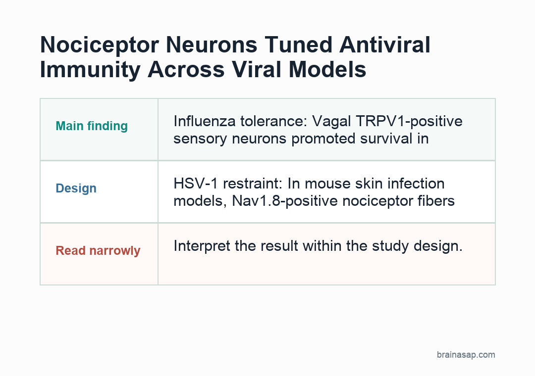
- HSV-1 restraint: In mouse skin infection models, Nav1.8-positive nociceptor fibers restrained neutrophil-heavy inflammation while supporting dendritic-cell priming of virus-specific CD8-positive T cells.
- Influenza tolerance: Vagal TRPV1-positive sensory neurons promoted survival in influenza models without clearly lowering viral load, mainly by limiting damaging lung myeloid inflammation.
- CGRP split role: Calcitonin gene-related peptide (CGRP) supported antiviral T helper 1 differentiation in acute LCMV and antibody responses during influenza, but the same mediator has different effects in other tissues.
- Chronic infection contrast: During chronic LCMV, sympathetic catecholamine signaling through ADRB1 deepened CD8-positive T cell exhaustion, showing that neural input is not uniformly protective.
Source: Journal of Neuroinflammation (2026) | Thomas et al.
Nociceptor neurons are usually introduced as pain-sensing fibers.
Thomas et al. review a more complicated role: these neurons sit in skin, lung, lymph nodes, sensory ganglia, and other tissues where viral infection and immune activation are happening.
The review frames pain fibers as local immune modulators. Their effect depends on the receptor, tissue, virus, and immune cell involved.
Nociceptors Can Detect Viral Cues Without Being Infected
Nociceptors can respond to infection even when a virus is not productively replicating inside the neuron. They express pattern-recognition receptors, including Toll-like receptors (TLRs) and RIG-I-like receptors, that can sense viral nucleic acids or infection-associated damage cues.
A viral illness can activate nociceptors through several routes at once. A neuron may detect viral material directly, respond to cytokines released by immune cells, or become sensitized by damage-associated molecular patterns from infected tissue.
- TLR4 and TRPV1: Damage cues and viral envelope-related inputs can lower the activation threshold of TRPV1, a heat- and irritant-sensitive channel on many nociceptors.
- TLR7 and TRPA1: Single-stranded RNA-related inputs can increase neuronal firing through TRPA1-linked pathways.
- TLR3 and TLR9: Double-stranded RNA and CpG DNA sensing can increase TRPV1 sensitivity and promote inflammatory mediator expression in sensory neurons.
- Type I interferon: IFN-alpha/beta receptor signaling can sensitize nociceptors through JAK1, TYK2, MNK, and eIF4E pathways.
Once activated, these neurons can release CGRP, substance P, and other mediators. Those outputs can alter blood-vessel tone, leukocyte movement, dendritic-cell behavior, B cell responses, and T cell polarization.
HSV-1 Models Linked Pain Fibers to Less Tissue Damage
Herpes simplex virus type 1 is a useful model because it infects barrier tissues and can enter sensory neurons. In cutaneous HSV-1 infection, the review describes mouse studies where broad Nav1.8-positive afferents helped limit excessive skin inflammation.
When those fibers were genetically removed, infected skin showed more neutrophil recruitment, higher inflammatory cytokine and chemokine production, and larger necrotic lesions. Viral clearance was not the main difference; immune dysregulation was.
- Nav1.8 fibers: Their absence increased neutrophil-heavy inflammation and reduced dendritic-cell priming of virus-specific CD8-positive T cells.
- TRPV1 neurons: This subset helped limit skin lesions through substance P signaling to neutrophils through MRGPRA1.
- GINIP neurons: Another sensory subset released TAFA4, which supported IL-10-dependent resolution of inflammation in dorsal root ganglia after viral clearance.
- Wounded skin: TRPV1-positive cutaneous nociceptors supported IL-27-dependent antiviral protein induction, including OAS-family antiviral genes.
In these models, selected sensory neurons promoted disease tolerance, meaning less tissue injury during infection, rather than simply killing more virus.
Influenza Circuits Split by Anatomy and Neuron Type
Influenza A virus shows why whole-class claims about nociceptors can fail. The review describes protective vagal and pulmonary sensory circuits, but also notes that other sensory subsets can worsen sickness behavior and survival in the same disease area.
In one lung-focused model, ablation of TRPV1-positive vagal neurons reduced survival after IAV infection without clearly reducing viral load. The affected mice had more lung damage, more neutrophils and monocyte-derived macrophages, and altered interferon-linked myeloid responses.
- Lung-vagal pathway: Pulmonary vagal sensory neurons helped maintain interferon-competent immune responses and limited damaging inflammation.
- Spleen pathway: TRPV1-positive sensory neurons supported germinal-center B cells, plasma-cell output, and virus-specific antibody production through CGRP signaling.
- Upper-airway pathway: GABRA1-positive glossopharyngeal sensory neurons responded to prostaglandin E2 through EP3 receptors and promoted sickness behaviors that reduced survival in a mouse H1N1 model.
The same virus therefore engaged several neural routes with different consequences. This context dependence is part of the biology the review asks researchers to map more precisely.
CGRP Helped Acute LCMV Immunity but Neural Stress Worsened Chronic Exhaustion
LCMV, an experimental arenavirus model, illustrates how infection stage changes the meaning of neural input. In acute LCMV Armstrong infection, neuronal CGRP signaling pushed naive T cells toward T helper 1 differentiation through CALCRL-RAMP3 receptor engagement.
That acute pathway supported IFN-gamma production, larger antiviral effector pools, and better viral control in the reviewed mouse work. The mechanism tied neuronal peptide release to a defined T cell receptor complex rather than a vague stress response.
- Acute LCMV: CGRP increased cAMP, CREB, ATF3, STAT1 activity, and IFN-gamma-linked T helper 1 differentiation.
- Chronic LCMV: Exhausted CD8-positive T cells localized near sympathetic axons and upregulated ADRB1, the beta-1 adrenergic receptor.
- ADRB1 blockade: Reducing catecholamine input lowered PD-1 and TIM-3 expression and improved T cell proliferation and cytokine production in chronic infection experiments.
This contrast is the review’s clearest warning for translation. A neural pathway that helps during acute viral control may not help during chronic antigen exposure, and a pain- or migraine-related target may also participate in host defense.
Translation Needs Circuit-Resolved Human Validation
The review is mostly a synthesis of animal and mechanistic studies, not a clinical treatment guide. Species differences, broad ablation methods, overlapping neuron markers, and pleiotropic mediators make direct therapeutic conclusions risky.
That caution is especially relevant for CGRP-targeting drugs. Blocking CGRP can be clinically useful in migraine care, but the reviewed biology suggests CGRP also supports selected antiviral T cell and antibody responses in some settings.
- Direct infection versus activation: Viral RNA in ganglia does not always prove productive infection of nociceptors.
- Subset identity: Nav1.8, TRPV1, GINIP, and GABRA1 labels capture overlapping but biologically different neuron populations.
- Tissue context: Skin, lung, spleen, ganglia, lymph node, and central nervous system circuits do not have interchangeable immune outputs.
- Human relevance: HSV entry into human sensory neurons and CGRP effects on human Langerhans cells support plausibility, but intervention-level evidence is still limited.
The review gives researchers a circuit map to test. Antiviral immunity is not only an immune-cell program; it is also shaped by local neural circuits that determine whether inflammation clears infection, damages tissue, supports antibodies, or reinforces exhaustion.
Citation: DOI: 10.1186/s12974-026-03817-z. Thomas et al. Nociceptor neurons shape antiviral immunity. Journal of Neuroinflammation. 2026;23:137.
Study Design: Narrative review of neuro-immune antiviral circuits across sensory neurons, autonomic inputs, viral infection models, and selected human-relevant findings.
Sample/Model: Review article; no new participant, animal, or cell dataset was generated by the authors.
Key Statistic: The review centers on circuit-defined findings including Nav1.8-positive HSV-1 inflammation restraint, TRPV1-positive influenza disease tolerance, CGRP-supported LCMV Th1 differentiation, and ADRB1-linked T cell exhaustion in chronic infection.
Caveat: Most evidence comes from animal or mechanistic models, so therapeutic targeting needs human validation at the level of specific neuron subsets, receptors, tissues, and infection stages.