TL;DR: A 2026 computational study in PLOS One used GEO Alzheimer’s datasets, 130 machine-learning models, and molecular simulations to identify eight candidate genes that may sit in a trichostatin A-related Alzheimer’s disease network.
Key Findings
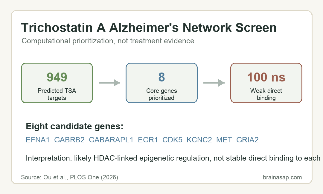
- 949 predicted TSA targets: The analysis started with predicted human protein targets for trichostatin A, a histone deacetylase inhibitor.
- Five GEO datasets: Researchers used two Alzheimer’s training datasets, two validation datasets, and one single-cell dataset from the Gene Expression Omnibus.
- Eight core genes: Machine-learning and network analyses highlighted EFNA1, GABRB2, GABARAPL1, EGR1, CDK5, KCNC2, MET, and GRIA2.
- Weak direct binding: 100 ns molecular dynamics simulations suggested trichostatin A did not stably bind key candidates such as CDK5 and GABRB2.
- Experimental gap: The paper’s own caveat is central: all findings are in silico and require cell, animal, and tissue-level validation.
Source: PLOS One (2026) | Ou et al.
Trichostatin A is a histone deacetylase inhibitor, meaning it can alter gene expression by changing acetylation patterns rather than acting like a single receptor-blocking drug.
Alzheimer’s disease is therefore a plausible but difficult target for computational work. The disease involves amyloid, tau, synaptic dysfunction, immune signaling, and cell-type-specific brain changes, so a one-target mechanism is unlikely to explain much on its own.
Machine Learning Screened Trichostatin A Targets Against Alzheimer’s Data
The researchers first predicted 949 potential trichostatin A targets using ChEMBL, SwissTargetPrediction, and PharmMapper. They then compared those targets with Alzheimer’s-related genes from public transcriptomic datasets.
The dataset design was layered rather than single-sample. Training used GSE122063 and GSE132903, validation used GSE44771 and GSE109887, and cell-type analysis used GSE161045.
The sample structure included several data levels:
- Training cohort one: 56 Alzheimer’s patients and 44 healthy controls.
- Training cohort two: 97 Alzheimer’s patients and 98 healthy controls.
- Validation cohorts: 129 Alzheimer’s patients with 101 controls, and 46 Alzheimer’s patients with 32 controls.
- Single-cell dataset: Four Alzheimer’s samples and four control samples.
After differential-expression analysis and co-expression-network analysis, the team used 130 machine-learning model combinations to narrow candidate genes. The aim was diagnostic and mechanistic prioritization, not a clinical drug trial.
Eight Core Genes Clustered Around Synapses, Tau, and Immune Signaling
The final candidate set contained EFNA1, GABRB2, GABARAPL1, EGR1, CDK5, KCNC2, MET, and GRIA2. The paper framed them as a regulatory network that could connect trichostatin A biology with Alzheimer’s disease processes.
Several genes map onto recognizable Alzheimer’s-relevant systems. CDK5 is tied to tau phosphorylation biology, while GABRB2 and KCNC2 point toward inhibitory signaling and neuronal excitability.
GRIA2 is part of AMPA glutamate receptor biology, and GABARAPL1 is linked to autophagy.
The expression pattern was not symmetric. The authors described broad downregulation of several putatively protective genes and distinct upregulation of EFNA1, which they interpreted as a possible pathological node in immune-neural crosstalk.
Molecular Dynamics Weakened the Direct-Binding Claim
Molecular docking initially suggested that trichostatin A could bind several candidate proteins. The reported docking energies included -9.7 kcal/mol for GABRB2, -8.9 kcal/mol for CDK5, and -8.6 kcal/mol for MET.
The stronger test came afterward. In 100 ns molecular dynamics simulations, the trichostatin A-CDK5 and trichostatin A-GABRB2 complexes did not show stable direct binding.
The reported binding energies after simulation were weak, including dG = -1.163 kcal/mol for CDK5 and -0.017 kcal/mol for GABRB2.
That result changed the interpretation. Instead of proposing that trichostatin A directly locks onto these Alzheimer’s-linked proteins, the analysis points back to histone deacetylase biology:
- HDAC1-3: Class I histone deacetylases that can regulate gene transcription.
- HDAC6: A cytoplasmic histone deacetylase linked in the paper to autophagy and protein-clearance hypotheses.
- Gene-network balance: A proposed shift in expression across tau, synaptic, and immune-related genes.
This is a more cautious mechanism. It treats trichostatin A as an epigenetic regulator that might reshape a network, not as a direct inhibitor of each candidate Alzheimer’s protein.
Single-Cell Analysis Placed Genes in Different Brain Cell Types
The single-cell part of the analysis used GSE161045 to ask where the candidate genes appeared across brain cell populations. The paper annotated astrocytes, endothelial cells or pericytes, microglia, neurons, oligodendrocytes, and oligodendrocyte precursor cells.
Cell-type placement added biological context to the gene list:
- Neurons: CDK5, GRIA2, and KCNC2 were described as neuron-enriched or closely tied to neuronal function.
- Oligodendrocytes: MET was reported as highly expressed in oligodendrocytes, suggesting a myelin-related angle.
- Vascular and glial cells: EFNA1 expression appeared across endothelial cells, pericytes, oligodendrocytes, and astrocytes.
The analysis also linked EFNA1 with immune-cell infiltration patterns, including plasma cells and CD8+ T cells.
Within the model, EFNA1 became a possible bridge between inflammation and neurodegeneration, but the paper did not prove that bridge experimentally.
The Alzheimer’s Claim Is a Prioritization Map, Not Treatment Evidence
The most important reader safeguard is the study design. This was a computational pipeline, not a patient trial, not a mouse treatment experiment, and not a cell-culture validation study.
The candidate genes, immune-neural crosstalk model, and trichostatin A mechanism all still lack experimental evidence from cell and animal models. The paper proposed future CRISPR experiments and trichostatin A intervention studies in Alzheimer’s mouse models.
For now, the useful output is a ranked hypothesis map. It says which genes and pathways might deserve experimental testing if researchers want to understand how histone deacetylase inhibition could intersect with Alzheimer’s pathology.
The prioritization still has value because Alzheimer’s drug discovery often fails when a plausible mechanism looks strong in one evidence layer but does not survive translation.
A computational screen can organize the next experiments, but it cannot substitute for them.
Citation: DOI: 10.1371/journal.pone.0347532. Ou et al. Deciphering the molecular network of Trichostatin A in regulating Alzheimer’s disease screening of core genes and mechanistic investigation based on multidimensional bioinformatics and molecular simulation. PLOS One. 2026;21(4):e0347532.
Study Design: Computational bioinformatics, machine-learning, single-cell, docking, and molecular-dynamics analysis using public Alzheimer’s datasets.
Sample/Model: Five GEO datasets, including transcriptomic training and validation cohorts plus one single-cell Alzheimer’s dataset.
Key Statistic: 130 machine-learning model combinations prioritized eight core genes, while 100 ns simulations suggested weak direct binding between trichostatin A and CDK5 or GABRB2.
Caveat: All findings are in silico and require experimental validation before they can support Alzheimer’s treatment claims.