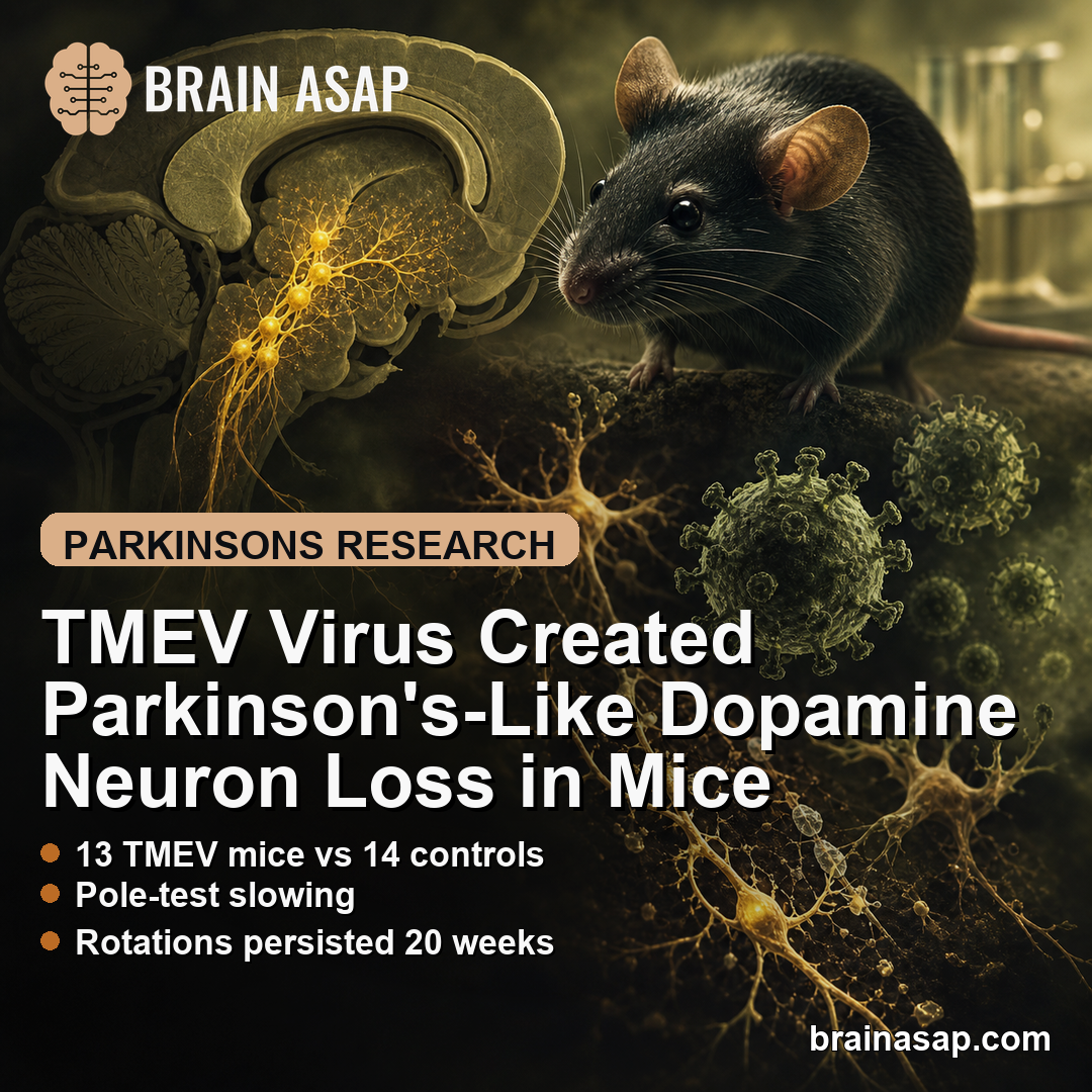
TL;DR: A 2026 study in Brain, Behavior, & Immunity – Health reported that Theiler’s murine encephalomyelitis virus (TMEV), a mouse neurotropic virus, infected substantia nigra dopamine neurons and produced Parkinson’s-like motor deficits in mice.
Key Findings
- 13 TMEV mice vs 14 controls: The main behavioral experiment compared unilateral TMEV injection into the substantia nigra with sham injection.
- Pole-test slowing: TMEV-injected mice took longer to descend at weeks 4, 16, and 20, with a significant injection effect for descent time (p = 0.0277).
- Rotations persisted 20 weeks: Apomorphine-induced contralateral rotations were higher at every measured post-injection timepoint from week 1 through week 20.
- Gait force changed: The left forelimb treadmill measure MINdAdT was lower after TMEV injection, with the week-12 difference reaching p = 0.0005.
- Dopamine-neuron staining fell: Tyrosine hydroxylase staining was lower on the injected substantia nigra side at 4 weeks (p = 0.041), while viral antigen was no longer detected then.
Source: Brain, Behavior, & Immunity – Health (2026) | Kang et al.
TMEV Tested a Viral Route to Parkinson’s-Like Dopamine Loss
Parkinson’s disease is defined in part by loss of dopamine-producing neurons in the substantia nigra pars compacta, a midbrain region that helps control movement. Most animal models recreate that loss with toxins or genetic manipulation.
This study tested a different route: direct infection with Theiler’s murine encephalomyelitis virus (TMEV). TMEV is a mouse virus already used in models of neuroinflammation, multiple sclerosis-like disease, and seizure biology.
The key question was narrow. If TMEV is placed into the substantia nigra, can it infect dopamine neurons, reduce tyrosine hydroxylase staining, and produce motor behavior resembling established unilateral Parkinson’s models?
Researchers used adult male C57BL/6J mice because this strain has a strong neuroinflammatory response after TMEV infection. The study does not show that common human viral infections cause Parkinson’s disease.
- Target region: substantia nigra pars compacta, the dopamine-neuron region damaged in Parkinson’s disease.
- Viral exposure: unilateral injection of TMEV into the right substantia nigra.
- Readouts: pole-test movement, apomorphine rotations, treadmill gait, viral staining, and tyrosine hydroxylase staining.
Pole-Test Slowing Suggested Parkinsonian Bradykinesia
The pole test measures how quickly a mouse turns and descends a vertical pole. In unilateral dopamine-lesion models, longer times are interpreted as impaired movement initiation or coordination.
After right-sided TMEV injection, mice generally took longer to perform the task than sham-injected controls. Descent time showed a significant overall injection effect, with p = 0.0277.
Post-hoc comparisons showed longer descent times at week 4, week 16, and week 20. Turning time also increased, reaching significance at weeks 4 and 16, with a week-20 trend.
The long follow-up is useful because a transient surgical effect would be expected to fade. Motor differences lasting out to 20 weeks fit better with ongoing circuit consequences after dopamine-neuron injury.
- Turn time: higher after TMEV injection at most timepoints, significant at weeks 4 and 16.
- Descend time: higher at every measured timepoint, significant at weeks 4, 16, and 20.
- Interpretation: the pattern resembles bradykinesia-like impairment in established dopamine-depletion models.
Apomorphine Rotations Pointed to Nigrostriatal Dopamine Injury
The apomorphine rotation test is a classic readout for one-sided nigrostriatal dopamine injury. When dopamine input falls on one side, postsynaptic dopamine receptors can become hypersensitive, and apomorphine can trigger turning toward the opposite side.
TMEV-injected mice showed more contralateral rotations than controls at every tested post-injection timepoint. The main injection effect was strong, with F(1,25) = 17.03 and p = 0.0004.
Week-by-week tests were significant at 1, 4, 8, 12, 16, and 20 weeks. The week-20 result was p = 0.0006, showing that the rotational behavior persisted to the end of follow-up.
The persistence strengthens the dopamine-lesion interpretation because the assay is tied to asymmetrical dopamine-system injury. It supports the idea that TMEV created a lasting unilateral lesion rather than a short-lived inflammatory sickness behavior.
Treadmill Gait and TH Staining Supported the Dopamine-Loss Model
The DigiGait treadmill system measured 48 gait parameters. Only one parameter, the maximal rate of change in paw area during the propulsion phase, clearly separated the groups.
That measure, called MINdAdT, reflects how quickly a limb unloads from the treadmill during the step cycle. In the left forelimb, MINdAdT was consistently lower after right-sided TMEV injection and reached p = 0.0005 at week 12.
The side of the effect made anatomical sense. The right brain helps control the left side of the body, so a right substantia nigra injury would be expected to affect left-sided movement.
Histology gave the behavioral result a tissue anchor. At 1 week, TMEV staining colocalized with tyrosine hydroxylase (TH)-positive dopamine neurons in the substantia nigra.
At 4 weeks, TH fluorescent intensity was lower on the injected side than the uninjected side in TMEV mice. The un-injected/injected side intensity ratio was higher than in controls, with p = 0.041.
- Virus location: TMEV colocalized with TH-positive substantia nigra neurons at 1 week.
- Neuron marker loss: TH staining was reduced on the injected side at 4 weeks.
- Virus clearance: viral antigen was no longer detected at the 4-week histology timepoint.
Viral Parkinson’s Models May Help Study Neuroinflammation
The main value of this work is model development. Toxin models such as 6-OHDA and MPTP can reproduce dopamine-neuron loss, but they do not start from an infection-centered inflammatory trigger.
A TMEV-based Parkinson’s model could help researchers ask how viral infection, host immune response, and dopamine-neuron vulnerability interact. That is different from claiming that TMEV is relevant to human exposure, since TMEV is a mouse pathogen.
Several limits remain before the model can be treated as established:
- Artificial route: the virus was injected directly into the substantia nigra rather than acquired naturally.
- Male mouse sample: the main experiment used adult male C57BL/6J mice, so sex and strain effects need more work.
- Model validation: additional studies should compare inflammation, alpha-synuclein biology, dopamine chemistry, and treatment response against standard Parkinson’s models.
Even with those limits, the result adds a useful tool. It shows that an infection-centered mouse experiment can reproduce several dopamine-lesion readouts that Parkinson’s researchers already know how to measure.
Citation: DOI: 10.1016/j.bbih.2026.101230. Kang et al. Theiler’s murine encephalomyelitis virus as the infectious agent for a virally induced mouse model of Parkinson’s disease. Brain, Behavior, & Immunity – Health. 2026;54:101230.
Study Design: Mouse disease-model study using unilateral TMEV injection into the substantia nigra with behavioral and histology readouts.
Sample/Model: Adult male C57BL/6J mice, including a main behavioral comparison of 13 TMEV-injected mice and 14 controls.
Key Statistic: Apomorphine-induced contralateral rotations were higher after TMEV injection at every measured post-injection timepoint through week 20.
Caveat: Direct-injection mouse model; it does not show that human viral infections cause Parkinson’s disease.