TL;DR: A 2026 Human Brain Mapping study combined PET receptor imaging with speech functional MRI, or fMRI, in healthy adults and found that GABA signaling and striatal dopamine release interacted with brain activity during human speech control.
Key Findings
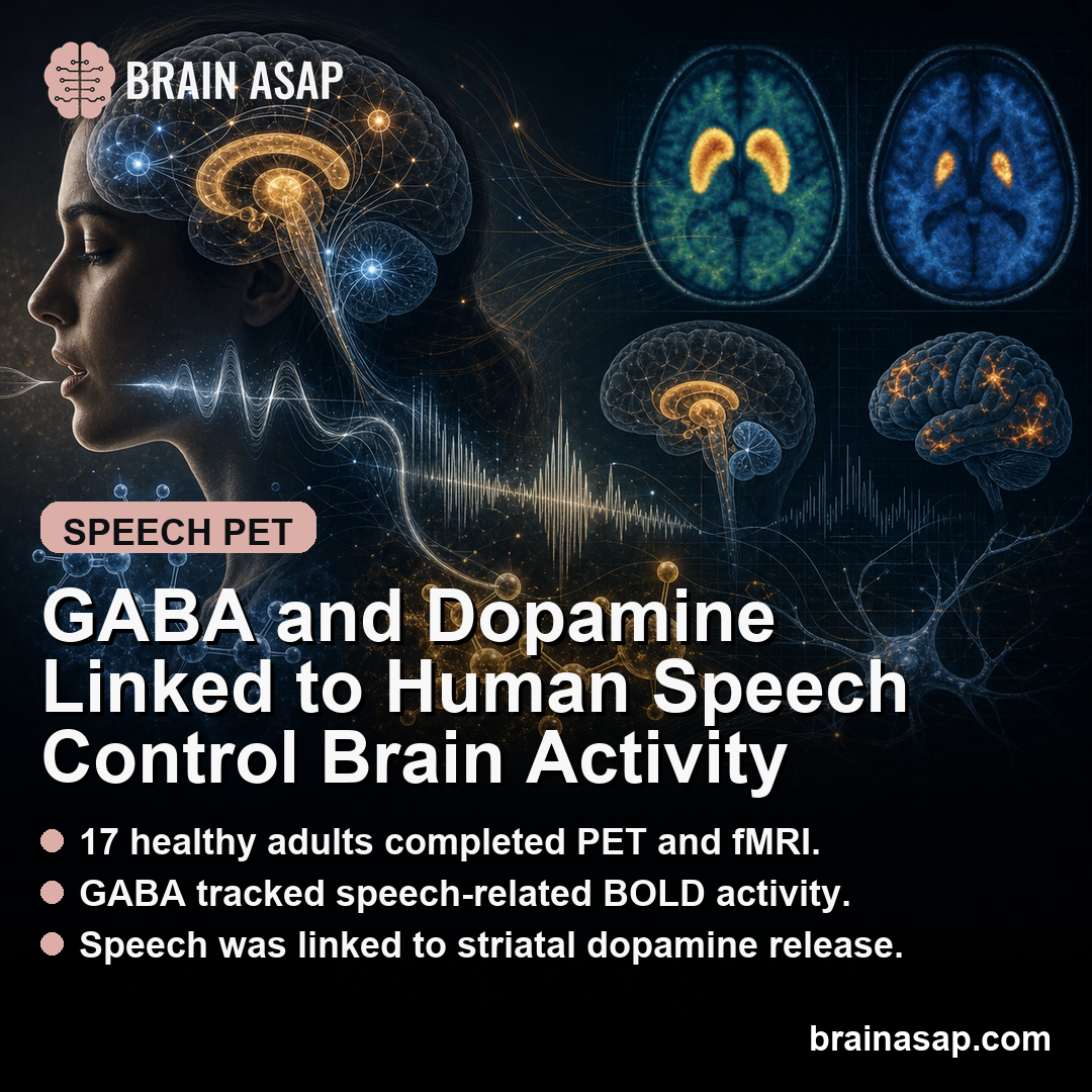
- PET-fMRI sample: The study included 17 right-handed volunteers, with mean age about 53 years.
- Receptor tracers separated systems: Researchers measured GABAA receptor availability with [11C]flumazenil and dopamine-related binding with [11C]raclopride.
- GABA tracked speech BOLD signals: GABAA receptor availability correlated with speech-related BOLD activity in regions including SMA, right inferior frontal gyrus, putamen, and cerebellum.
- Dopamine release occurred in striatum: Speech was associated with dopamine-related raclopride binding changes in left caudate and putamen.
- Interaction mattered most: GABAergic and dopaminergic measures jointly modulated activity in left associative caudate and sensorimotor putamen.
Source: Human Brain Mapping (2026) | Battistella et al.
PET and fMRI Linked Neurochemistry to Human Speech Control
Fluent speech requires fast coordination across motor planning, auditory feedback, basal ganglia circuits, and cortical control regions.
This study tested how GABA, the brain’s main inhibitory neurotransmitter, and dopamine, a major striatal neuromodulator, relate to the brain activity that supports speech.
The researchers combined positron emission tomography, or PET, with functional MRI.
PET measured receptor and neurotransmitter-related biology, while fMRI measured task-related blood oxygenation level-dependent, or BOLD, signals during speech.
The combination let the study connect molecular signaling to network-level activity.
That multimodal design is the main strength.
A standard fMRI study can show which regions are active during speech, but it cannot directly say how GABAergic and dopaminergic systems are involved.
PET adds a slower but more chemically specific view of the same system.
The approach also fits a broader question in speech neuroscience.
Speech symptoms appear in disorders that affect the cortex, basal ganglia, cerebellum, or neurotransmitter systems.
A molecular-circuit bridge can help researchers move beyond region lists toward mechanisms that may explain why speech timing and fluency are vulnerable.
Healthy Volunteers Completed Speech Imaging With PET Tracers
The study included 17 healthy right-handed volunteers, with mean age 53.2 +/- 10.1 years; 13 were female. Participants completed speech-related fMRI and PET imaging with two radiotracers.
[11C]flumazenil was used to estimate GABAA receptor availability. GABAA receptors mediate fast inhibitory signaling, which helps shape timing and gain across speech-motor circuits.
[11C]raclopride was used to assess dopamine D2/3 receptor-related binding, a standard way to infer dopamine release during a task when binding changes from rest to activation.
Researchers tested whether GABA receptor availability and dopamine release during speech aligned with speech-network activation.
This is not the same as testing a medication or stimulation protocol.
Receptor availability can reflect a person’s neurochemical baseline, while task-related dopamine release captures a dynamic response during speech.
The study put those two measurements into the same analytic frame.
GABAA Receptor Availability Correlated With Speech BOLD Activity
GABAA receptor availability showed strong positive correlations with speech-related BOLD responses in several regions.
The supplementary motor area, or SMA, had one of the clearest relationships, with Rs = 0.84 and p = 2e-5.
Positive correlations also appeared in the right inferior frontal gyrus, including pars opercularis (Rs = 0.65, p = 0.005) and pars triangularis (Rs = 0.66, p = 0.004).
Additional associations were reported in left parietal lobules, right superior temporal gyrus, left putamen, and cerebellum.
These regions fit speech control biology:
- SMA: Motor planning, initiation, and internally guided sequencing.
- Inferior frontal cortex: Speech production and language-motor control.
- Superior temporal and parietal regions: Auditory and sensorimotor integration.
- Putamen and cerebellum: Timing, sequencing, and motor execution.
The study also found a negative correlation in left inferior and middle frontal gyri, with Rs = -0.74 and p = 7e-4.
GABA-BOLD relationships therefore differed by region rather than following one global direction across the brain. Dopamine-related PET findings centered on the striatum.
During speech, the researchers reported raclopride binding changes in left anterior caudate, posterior caudate, and putamen. The reported binding reductions included approximately -8.6%, -9.5%, and -6.3% in those striatal regions.
In raclopride PET studies, lower binding during a task can be interpreted as greater endogenous dopamine competing with the tracer.
The finding is therefore consistent with dopamine release during speech, especially in striatal circuits involved in action selection and motor control.
The left striatal emphasis also makes biological sense.
Speech is a learned, precisely sequenced motor behavior, and basal ganglia circuits help regulate initiation, timing, and fluent execution.
Dopamine is central to those circuits, including in movement disorders where speech can become slow, effortful, or dysfluent.
The result should be interpreted as a task-related signal, not as a simple dopamine level.
Raclopride PET detects changes in binding potential, and the usual interpretation depends on competition between the tracer and endogenous dopamine.
The method is informative but indirect.
GABA and Dopamine Interacted in Caudate and Putamen
The most important result was not simply that GABA and dopamine each related to speech activity. The study found evidence that they interacted.
Neural activity during speech was modulated by the interaction between GABAA receptor availability and striatal dopamine release in two left striatal regions:
- Associative caudate: R = -0.63, p = 0.007.
- Sensorimotor putamen: R = -0.67, p = 0.003.
That points to a circuit-level interpretation. Speech control may depend on the balance between inhibitory cortical signaling and dopamine-mediated striatal modulation, rather than on either system in isolation.
The caudate and putamen are well positioned for that interaction because they connect motor planning, action selection, and learned speech sequences.
The interaction finding also helps explain why speech disorders can be hard to localize to one neurotransmitter or one region.
A person could have cortical inhibition, striatal dopamine signaling, or network timing differences that only become visible during active speech production.
This study does not resolve those disease questions, but it gives them a sharper circuit model.
Fluent speech does not appear to depend on a single chemical switch.
The PET-fMRI results suggest coordination between inhibitory control and dopamine-sensitive action pathways, which fits a behavior that has to be fast, precise, and flexible.
Resting-state correlations were much more limited, appearing mainly in visual cortex and cerebellar lobule VI. The speech task therefore appeared to reveal neurochemical relationships that were not obvious during rest.
Small PET-fMRI Study Limits the Clinical Claim
The strongest claim is mechanistic.
In healthy adults, GABAA receptor availability and speech-related dopamine release were linked to neural activity in speech-control circuits.
The study does not prove that changing either neurotransmitter system would improve speech.
Several limits constrain the claim:
- Small sample: PET-fMRI studies are expensive and intensive, but 17 participants limits precision.
- Healthy adults: The findings do not directly establish disease mechanisms in stuttering, Parkinson’s disease, dystonia, or aphasia.
- Correlational design: Receptor availability, dopamine release, and BOLD activity were associated, not experimentally manipulated.
- Task specificity: Results may depend on the speech task and imaging model used in the study.
The paper still gives speech neuroscience a valuable bridge between molecules and circuits.
It suggests that human speech control is shaped by a coordinated GABA-dopamine system, especially in cortico-striatal pathways that help transform planned speech into fluent movement.
Citation: DOI: 10.1002/hbm.70531. Battistella et al. Linking GABAergic and dopaminergic neurotransmission: Effects on neural activity during human speech control. Human Brain Mapping. 2026.
Study Design: PET receptor imaging combined with speech-task functional MRI.
Sample/Model: 17 healthy adults completed PET-fMRI speech-control imaging.
Key Statistic: GABAA receptor availability and striatal dopamine release aligned with speech-network activation.
Caveat: Small healthy-volunteer PET-fMRI data cannot establish clinical speech-disorder mechanisms.