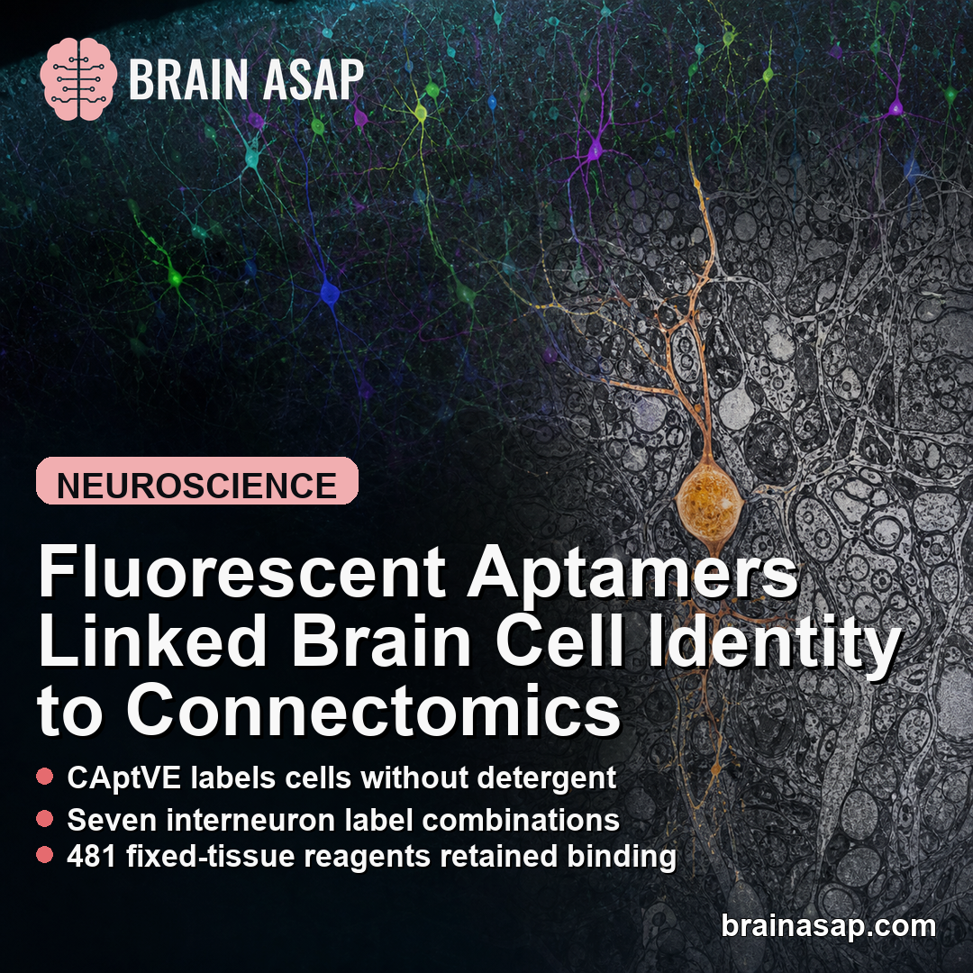
TL;DR: A 2026 Nature Communications paper introduced CAptVE, a fluorescent aptamer method that labels molecular features in mouse brain tissue while preserving ultrastructure for electron-microscopy connectomics.
Key Findings
- Detergent-free labeling: CAptVE used slow off-rate modified aptamers to label brain cells without detergent permeabilization that can disrupt ultrastructure.
- Three fixation steps: The optimized pipeline balanced fluorescence labeling with electron-microscopy preservation through staged fixation.
- Seven label combinations: SST, CR, and NPY labeling identified all seven possible molecular combinations in mouse cortical interneurons.
- 451 labeled cells: One analysis found a rare SST-/CR+/NPY+ type represented by only 2 of 451 labeled cells.
- 481 fixed-tissue reagents: After paraformaldehyde fixation, 481 SOMAmer reagents retained enough binding for fixed mouse brain tissue.
Source: Nature Communications (2026) | Lu et al.
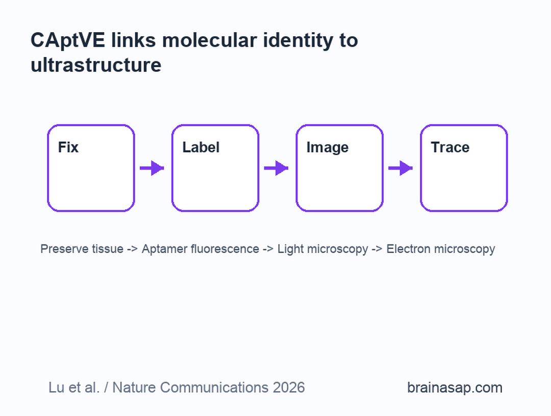
CAptVE stands for an aptamer-based approach to correlated light and electron microscopy. The goal is to connect two kinds of information that brain researchers often need separately: molecular cell identity and ultrastructural circuit detail.
That problem is central to connectomics. Electron microscopy can trace tiny neuronal processes and synapses, but it is harder to know exactly which molecular cell type each reconstructed neuron belongs to.
Fluorescent Aptamers Added Molecular Labels to Brain Ultrastructure
Aptamers are short nucleic-acid binders selected to recognize protein targets. The paper used slow off-rate modified aptamers, also called SOMAmer reagents, as fluorescent labels for fixed brain tissue.
Traditional antibody staining often needs detergent to let large antibodies enter tissue. Detergent can damage membranes and extracellular space, which are exactly the structures electron microscopy needs to preserve.
- Molecular identity: Fluorescent labels mark proteins associated with specific brain-cell classes.
- Ultrastructure: Electron microscopy preserves membranes, synapses, mitochondria, and fine cellular architecture.
- Connectomics use: Overlaying the two lets researchers connect cell-type labels with reconstructed circuits.
The CAptVE method tries to make those layers compatible. It labels cells first, then prepares the same tissue for high-resolution electron microscopy.
Fixation Had to Preserve Both Binding and Structure
The methods problem was delicate. Mild fixation preserved aptamer binding but did not preserve ultrastructure well enough. Overnight fixation preserved structure but weakened aptamer binding.
The final protocol used 4% paraformaldehyde for 3 hours, then additional fixation steps after aptamer binding and before electron-microscopy preparation. Most processing was kept at 4°C before osmium fixation to protect membrane integrity.
- Initial fixation: Tissue was fixed strongly enough to remain usable but not so strongly that epitopes disappeared.
- Aptamer staining: SOMAmer reagents were applied before deeper post-fixation locked the tissue down.
- EM preparation: After fluorescence imaging, tissue was prepared for electron microscopy with ultrastructure preserved.
Researchers checked sensitive structures such as mitochondria, plasma membranes, and synapses. They reported that the damaging artifacts expected from poor fixation were not observed in the assessed samples.
SST, CR, and NPY Labels Split Cortical Interneurons
The team tested volumetric correlated light and electron microscopy using aptamers against calretinin (CR) and neuropeptide Y (NPY), alongside SST-positive interneuron labeling.
In mouse somatosensory cortex, fluorescence identified all seven possible combinations of SST, CR, and NPY expression. The abundance of these combinations varied by cortical layer.
- L1 pattern: SST-/CR-/NPY+ cells were most abundant in layer 1.
- L2/3 and L4 pattern: SST-/CR+/NPY- cells predominated in layers 2/3 and 4.
- L5 and L6 pattern: SST+/CR-/NPY- cells were most abundant in layers 5 and 6.
The electron-microscopy analysis then linked those molecular labels with cell morphology. For example, CR-only neurons had more nuclear infoldings, with a surface-area-to-volume ratio of 1.57 compared with 0.97 to 1.26 in other cell types.
Proteome-Scale Screening Found Fixed-Tissue Brain Labels
The study also explored a larger SOMAmer library. Out of more than 7,000 reagents available at the time, 1,132 specifically bound protein targets in lysed frozen mouse brain using a 3-times-background threshold.
Fixation reduced binding for most reagents. After paraformaldehyde treatment, 481 reagents still maintained enough binding in fixed mouse brain tissue.
- Astrocyte labels: PRDX6 labeled astrocyte somata and vascular endfeet, while GFAP mainly localized to astrocytic processes.
- Neuron labels: PENK, NEUG, PRKCG, ELAVL2, and NPTXR labeled distinct neuronal populations or processes.
- Glia and myelin labels: QDPR marked a subset of oligodendrocytes, while MAG was limited to myelin sheath.
This is why the method could scale. If many aptamers remain useful after fixation, researchers can add molecular labels beyond a small handpicked antibody panel.
CAptVE Is a Research Tool, Not a Clinical Test
The study was performed in mouse brain tissue and focused on technical validation. It does not diagnose disease, measure a patient’s brain health, or provide a clinical biomarker test.
Its importance is upstream. Brain atlases increasingly define cell types by transcriptomics and proteomics, while connectomics defines circuits by structure. CAptVE offers a way to bring those maps closer together.
The method also helps explain why a short molecular label is not always enough. Cells with the same sparse marker combination could still differ in dendritic shape, nuclear structure, or protein-expression level, so structure and molecular identity need to be read together.
- Research strength: The method preserves ultrastructure while adding molecular identity.
- Current limit: Label multiplexing needs to expand, and aptamer performance depends on fixation-sensitive binding.
- Future use: Larger aptamer panels could help map how specific brain-cell types wire into circuits.
For neuroscience, the paper is a methods advance. It gives researchers another route toward seeing what a cell is, where it connects, and how its fine structure differs from neighboring cells.
That kind of integrated map could become especially useful when researchers compare normal circuits with models of brain disease, where cell-type vulnerability and synaptic wiring may change together.
Citation: DOI: 10.1038/s41467-026-72180-7. Lu et al. Probing molecular diversity and ultrastructure of brain cells with fluorescent aptamers. Nature Communications. 2026.
Study Design: Mouse brain methods study combining fluorescent aptamer labeling with volumetric correlated light and electron microscopy.
Sample/Model: Fixed mouse brain tissue, including GABAergic cortical interneuron and SOMAmer biomarker-labeling experiments.
Key Statistic: 481 SOMAmer reagents retained sufficient binding in fixed mouse brain tissue after paraformaldehyde treatment.
Caveat: The method is a research platform and needs broader multiplexing and validation before routine atlas-scale use.


