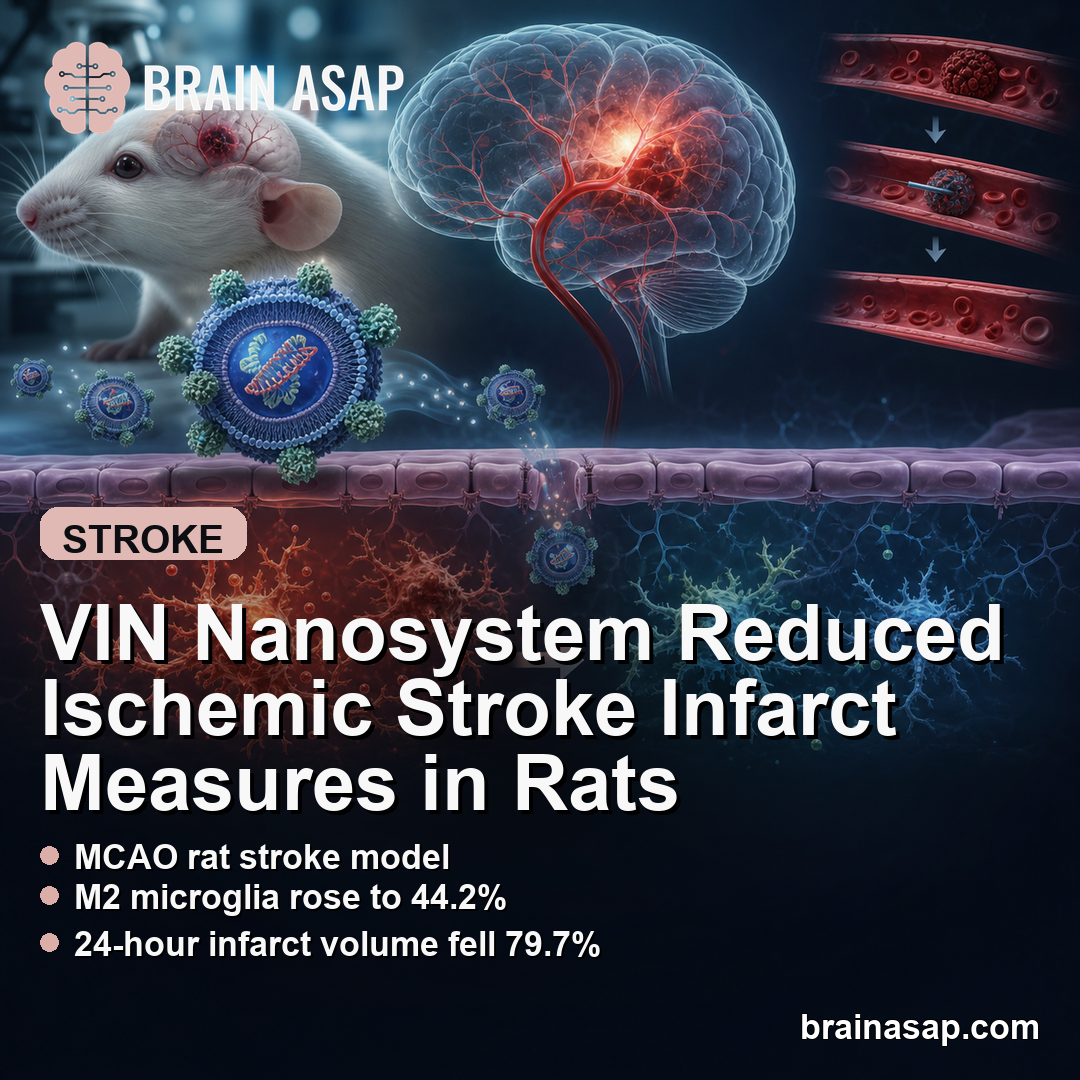
TL;DR: A 2026 rat and cell study in Journal of Materials Science: Materials in Medicine reported that an engineered VIN nanosystem shifted microglia toward an anti-inflammatory state and reduced ischemic-stroke infarct measures in MCAO rats.
Key Findings
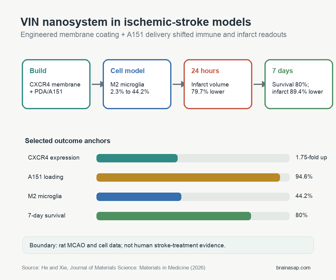
- CXCR4 increased 1.75-fold: Fe3O4 nanoparticle induction raised CXCR4 expression in bone-marrow mesenchymal stem cell membranes.
- A151 loading reached 94.6%: The polydopamine/Zn2+ system loaded the A151 oligonucleotide with reported efficiency of 94.6 ± 1.2%.
- M2 microglia rose to 44.2%: VIN treatment increased CD206-positive microglia from 2.3% to 44.2% in the cell model.
- 24-hour infarct volume fell: VIN produced a 79.7% infarct-volume reduction in the short-term rat MCAO model.
- Seven-day infarct volume fell 89.4%: In the long-term model, VIN increased survival to 80% and reduced infarct volume by approximately 89.4%.
Source: Journal of Materials Science: Materials in Medicine (2026) | He and Xie
Ischemic stroke damages brain tissue first through blocked blood flow, then through reperfusion injury when blood returns and immune signaling surges. This preclinical study tested a drug-delivery system built to target that post-stroke immune microenvironment.
The treatment was called a versatile immunosuppressive nanosystem, or VIN. It combined a stem-cell-membrane coating, CXCR4-targeting biology, polydopamine nanoparticles, zinc-mediated loading, and the A151 oligonucleotide.
VIN Combined CXCR4 Membrane Targeting With A151 Delivery
The researchers first induced bone-marrow mesenchymal stem cells with Fe3O4 nanoparticles, then extracted membranes with higher CXCR4 expression. CXCR4 is relevant because it can participate in homing toward ischemic injury signals.
The final VIN construct wrapped A151-loaded polydopamine nanoparticles with those engineered membranes. A151 was included because it can inhibit TLR9-related inflammatory signaling.
- Fe3O4 induction: Used to raise CXCR4 expression on the source cell membranes.
- Polydopamine core: Provided a nanoparticle scaffold with antioxidant and loading properties.
- Zn2+ bridge: Helped load A151 onto the polydopamine system.
- Cell-membrane coating: Intended to improve biocompatibility and lesion targeting.
This is a materials-and-neuroinflammation strategy, not a standard small-molecule stroke drug. The core question was whether the construct could reach the injured environment and quiet damaging immune activation.
CXCR4 Upregulation and A151 Loading Supported the Nanosystem Design
Characterization experiments supported the assembly logic. Fe3O4 nanoparticle exposure increased CXCR4 expression by 1.75-fold compared with control membranes.
Polydopamine and zinc also loaded the fluorescent A151 probe efficiently. The reported loading efficiency was 94.6 ± 1.2%, and the final VIN particle size was about 209.1 ± 15.6 nm.
- CXCR4 result: The membrane-targeting feature increased before nanosystem assembly.
- Loading result: A151 was efficiently associated with the nanoparticle system.
- Particle result: The larger final particle size supported membrane-coated VIN formation.
Transmission electron microscopy also showed a core-shell structure with a membrane coating about 14 nm thick. Those measurements matter because failed assembly would make later biological results much harder to interpret.
VIN Shifted Microglia Toward an Anti-Inflammatory Phenotype
Microglia are brain-resident immune cells that can amplify injury or support repair depending on activation state. The study measured CD16/32 as an M1-like pro-inflammatory marker and CD206 as an M2-like anti-inflammatory marker.
Lipopolysaccharide increased CD16/32-positive cells, consistent with pro-inflammatory activation. VIN treatment reduced CD16/32-positive cells and increased CD206-positive cells, with the M2-phenotype proportion rising from 2.3% to 44.2%.
- Pro-inflammatory side: VIN reduced CD16/32-positive microglia after inflammatory stimulation.
- Anti-inflammatory side: VIN increased CD206-positive microglia.
- Cytokine support: ELISA results showed higher IL-10 and Arg-1 with lower TNF-alpha and IL-6.
The cell data fit the proposed mechanism: A151 and the coated nanosystem appeared to steer microglial signaling away from inflammatory injury and toward a repair-associated profile.
Short-Term MCAO Rats Had Lower Infarct Volume After VIN
The in vivo testing used middle cerebral artery occlusion, or MCAO, a common rat model of ischemic stroke. In the short-term model, rats were assessed 24 hours after treatment.
VIN was compared with saline and several component controls, including A151 and membrane-coated constructs. The reported short-term infarct-volume reduction for VIN was 79.7% after therapy.
TUNEL staining also suggested less neuronal apoptosis in the VIN group. Immunofluorescence readouts showed lower MPO, Iba-1, GFAP, and 8-OHG signals, which the study interpreted as lower neutrophil infiltration, microglial activation, astrocyte activation, and oxidative stress.
- TTC staining: Used to quantify pale infarcted tissue.
- TUNEL staining: Used to identify apoptotic cells.
- 8-OHG signal: Used as an oxidative-stress marker.
These endpoints are preclinical tissue-readouts. They support neuroprotection in the model, but they do not show functional recovery in human stroke patients.
Seven-Day VIN Treatment Improved Survival and Infarct Measures
The long-term rat model used 4 VIN doses and evaluated animals over 7 days. Survival was 80% in the VIN group compared with 30% in saline-treated MCAO rats.
Body weight also recovered faster in VIN-treated rats. By day 7, the VIN group had returned near normal weight, while saline-treated rats continued to show poorer recovery.
TTC staining after 7 days showed saline-treated survivors with about 19.8% infarct volume. VIN treatment reduced infarct volume by approximately 89.4%, although the source notes survival bias as an issue in the long-term comparison.
- Survival: VIN-treated rats had higher seven-day survival than saline-treated MCAO rats.
- Weight recovery: VIN-treated rats regained weight earlier and more completely.
- Infarct size: Seven-day infarct volume was sharply lower after VIN treatment.
The survival-bias caveat is important. If many severe saline-treated rats died before day 7, the remaining saline group may underrepresent the worst injuries.
Preclinical Stroke Nanomedicine Still Faces Translation Barriers
VIN is promising as a proof-of-concept because it addresses several stroke barriers at once: targeting, blood-brain-barrier crossing, oxidative stress, and immune polarization. Combining all of those features also makes translation harder.
Human stroke treatment has tight time windows, variable clot locations, comorbid disease, and strong safety requirements. A complex membrane-coated nanosystem would need pharmacokinetics, biodistribution, toxicity, manufacturing, and large-animal validation before clinical testing.
- Model limit: MCAO rats are useful but do not reproduce the full diversity of human ischemic stroke.
- Timing limit: Treatment was delivered very early after embolization and reperfusion.
- Manufacturing limit: Cell-membrane-coated nanomedicines require reproducible quality control.
The preclinical message is still clear. In this source, VIN shifted microglial phenotype and reduced infarct-related injury markers in rat stroke models, making the immune microenvironment a plausible target for further stroke-nanomedicine work.
Citation: DOI: 10.1007/s10856-026-07014-5. He and Xie. Therapeutic effects of an engineered bionic decoy-integrated versatile immunosuppressive nanosystem based on an in vitro blood-brain barrier model in ischemic stroke. Journal of Materials Science: Materials in Medicine. 2026.
Study Design: Preclinical nanomedicine study using cell experiments plus short-term and seven-day MCAO rat ischemic-stroke models.
Sample/Model: Microglial cell assays and male Sprague-Dawley rat MCAO models, including 6 rats per group in the short-term model and 10 rats per group in the long-term model.
Key Statistic: VIN increased CD206-positive microglia from 2.3% to 44.2%, reduced short-term infarct volume by 79.7%, and reduced seven-day infarct volume by approximately 89.4%.
Caveat: These are cell and rat-model results; the source does not show clinical benefit, dosing safety, or manufacturing readiness in human stroke patients.