TL;DR: A 2026 study in Communications Biology found that restraint stress weakened medial prefrontal cortex (mPFC, a frontal brain region that helps organize social behavior) activity during social testing, while stress-activated amygdala and hypothalamic oxytocin inputs pushed the circuit toward inhibition.
Key Findings
- Two-hour acute restraint preserved basic sociability but disrupted social novelty preference, suggesting a short-term social-recognition problem.
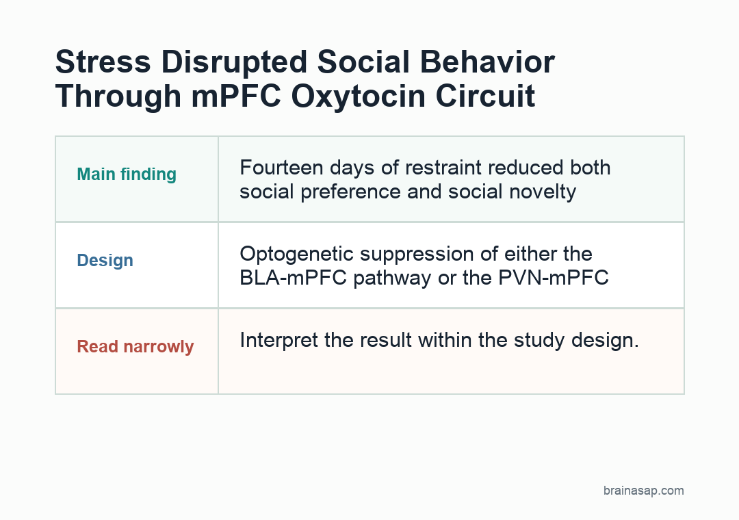
- Fourteen days of restraint reduced both social preference and social novelty preference, with preferred-stimulus interaction falling to about 8-16% of each session versus more than 20% in controls.
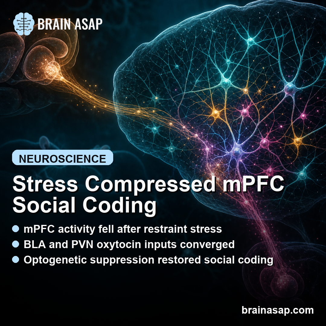
- mPFC calcium transient rates fell to about 70-80% of baseline after acute stress and nearly 50% of baseline after chronic stress.
- BLA and PVN inputs became stress-activated upstream pathways into the mPFC, with BLA meaning basolateral amygdala and PVN meaning paraventricular hypothalamus.
- Optogenetic suppression of either the BLA-mPFC pathway or the PVN-mPFC oxytocin pathway restored mPFC social coding and improved stressed mice’s social behavior.
Source: Communications Biology (2026) | Zhang et al.
Stress Changed Social Recognition Before It Changed Social Drive
Researchers used a repeated restraint model to separate short-term and longer-term stress effects in male C57BL/6 mice. The acute condition involved 2 hours of restraint.
The chronic condition involved 14 days of restraint, 2 hours per day. Mice were then tested in a three-chamber task that measures two related but separable behaviors.
- Social preference: whether the mouse spends more time near another mouse than near an object.
- Social novelty preference: whether the mouse spends more time near a new mouse than near a familiar mouse.
That distinction mattered. After acute stress, mice still preferred the social stimulus over the object, but they no longer clearly preferred the novel mouse over the familiar one.
In a separate two-trial test, acutely stressed mice still approached another mouse, but they failed to reduce sniffing when the same mouse was presented again. The pattern fits impaired social recognition, not simple social avoidance.
Chronic stress produced a broader phenotype. After 14 days, mice no longer showed the same social preference and also showed worse novelty preference.
Researchers reported that chronically stressed mice spent only 8-16% of each session interacting with preferred stimuli, compared with more than 20% in controls. Locomotion did not explain the finding because stressed and control mice traveled comparable distances during social testing.
mPFC Activity Lost Its Social-Discrimination Pattern
The medial prefrontal cortex, or mPFC, is often treated as a control hub for social behavior because it helps represent social cues, update context, and coordinate approach or recognition.
To watch that circuit directly, researchers expressed the calcium sensor GCaMP6f in excitatory prelimbic mPFC neurons and used miniscope imaging while mice performed the social task.
The imaging readout was not subtle. Researchers recorded 573 neurons at baseline, 612 neurons after acute stress, and 683 neurons after chronic stress.
During social testing, acute stress reduced mPFC calcium transient rates to roughly 70-80% of baseline. Chronic stress pushed the response down further, to nearly 50% of baseline.
The analysis also looked beyond average activity. Healthy mPFC ensembles normally separate overlapping social representations through pattern decorrelation, meaning different stimuli evoke distinguishable coactivity patterns across neurons.
Under stress, that separation weakened. After chronic stress, the difference in correlation-distribution width between preferred and nonpreferred stimuli almost collapsed toward zero.
Principal component analysis told the same story in a different format. Control mice maintained large distances between neural population states for different stimuli.
Acute stress compressed the representation of familiar versus novel social stimuli. Chronic stress compressed both social-versus-object and familiar-versus-novel representations. The weaker the neural separation, the worse the behavioral discrimination index.
Amygdala and Hypothalamus Converged on the Same Prefrontal Bottleneck
The next question was where the inhibitory pressure came from. The study focused on two stress-sensitive input systems that project into the prelimbic mPFC.
- BLA: the basolateral amygdala, a region involved in threat, salience, and emotional learning.
- PVN: the paraventricular hypothalamus, a stress-regulation hub that can release oxytocin and other neuropeptides.
Social testing after stress increased c-Fos, an activity marker, in both the BLA and PVN. The increase grew with stress duration and nearly doubled after 14 days of restraint.
At the same time, social-related c-Fos in the prelimbic mPFC fell, matching the calcium-imaging evidence for prefrontal hypoactivity.
Viral tracing showed that both upstream regions send projections into the prelimbic mPFC. The convergence matters because it gives stress two routes into the same social-coding circuit.
The BLA route appeared to recruit local GABAergic interneurons, which can inhibit nearby excitatory neurons. The PVN route was more peptide-specific: oxytocin-projecting neurons into the mPFC became unusually active after stress.
Suppressing Either Stress Input Restored Prefrontal Coding
The rescue experiments are the strongest part of the paper. Researchers used optogenetics, a method that can suppress genetically targeted neurons or axons with light, while also monitoring mPFC activity.
In stressed mice, suppressing BLA axons in the mPFC increased mPFC transient rates, restored pattern decorrelation, widened principal-component distances between stimuli, and brought social performance closer to baseline.
PVN input showed a related but more specific mechanism. Broad PVN-mPFC suppression helped acute-stressed mice and partially helped chronic-stressed mice.
When researchers targeted the PVN-mPFC oxytocin pathway, the behavioral rescue was stronger: social performance improved after both acute and chronic stress. Suppressing a comparison corticotropin-releasing hormone pathway did not produce the same improvement.
Additional pharmacology supported the oxytocin-interneuron model. Giving oxytocin before mild stress worsened social novelty preference and increased mPFC GABA-neuron responses.
Giving an oxytocin receptor antagonist after acute stress improved novelty preference and reduced GABAergic activity. In plain terms, stress-linked oxytocin input appeared to push the mPFC toward too much inhibition during social discrimination.
Mouse Circuit Evidence Stops Short of Treatment Rules
This is a mouse circuit study, so it does not say that blocking amygdala input or oxytocin signaling is a near-term treatment for human social problems after stress. The interventions were invasive, cell-type-specific laboratory manipulations.
The behavioral task also captures a narrow set of social approach and recognition behaviors, not the full range of human stress, trauma, depression, autism, or social withdrawal.
The study links stress exposure to a sequence that can be tested: BLA and PVN activity rise, inhibitory pressure inside mPFC increases, excitatory mPFC social coding becomes weaker, and social discrimination suffers.
The rescue experiments make the sequence more than a correlation because reducing either stress-activated input restored both neural coding and behavior in the stressed animals.
The finding narrows the question. Stress changed prefrontal coding, not only outward social behavior.
It compressed a prefrontal coding system that normally helps distinguish familiar, novel, social, and nonsocial cues. That kind of circuit-level explanation is useful because it names where stress changes the computation, not just the outward behavior.
Citation: DOI: 10.1038/s42003-026-10089-z. Study authors et al. Zhang et al. Medial prefrontal cortex neurons integrate amygdala and hypothalamic oxytocin signals to mediate stress-induced social alterations. Communications Biology . 2026.
Study Design: Mouse restraint-stress experiment with behavior testing, miniscope calcium imaging, viral tracing, optogenetic suppression, fiber photometry, and oxytocin pharmacology.
Sample/Model: Male C57BL/6 mice tested after acute 2-hour restraint or 14-day repeated restraint; mPFC imaging included 573 baseline neurons, 612 acute-stress neurons, and 683 chronic-stress neurons.
Key Statistic: mPFC calcium transient rates fell to about 70-80% of baseline after acute stress and nearly 50% after chronic stress.
Caveat: The findings come from invasive mouse circuit experiments and should not be treated as a direct clinical intervention strategy.