TL;DR: A 2026 study in Human Brain Mapping found that full-term neonates already showed stronger structural and functional connectivity between cortical gyri than between sulci, with brain structure-function coupling shifting toward decoupling around 41 weeks postmenstrual age.
Key Findings
- Neonatal imaging sample: Researchers analyzed full-term neonates from the developing Human Connectome Project scanned between 38.14 and 44.71 weeks postmenstrual age.
- 64 cortical ROIs: The analysis split 32 cortical regions into gyral and sulcal subregions, creating 64 regions of interest for connectivity mapping.
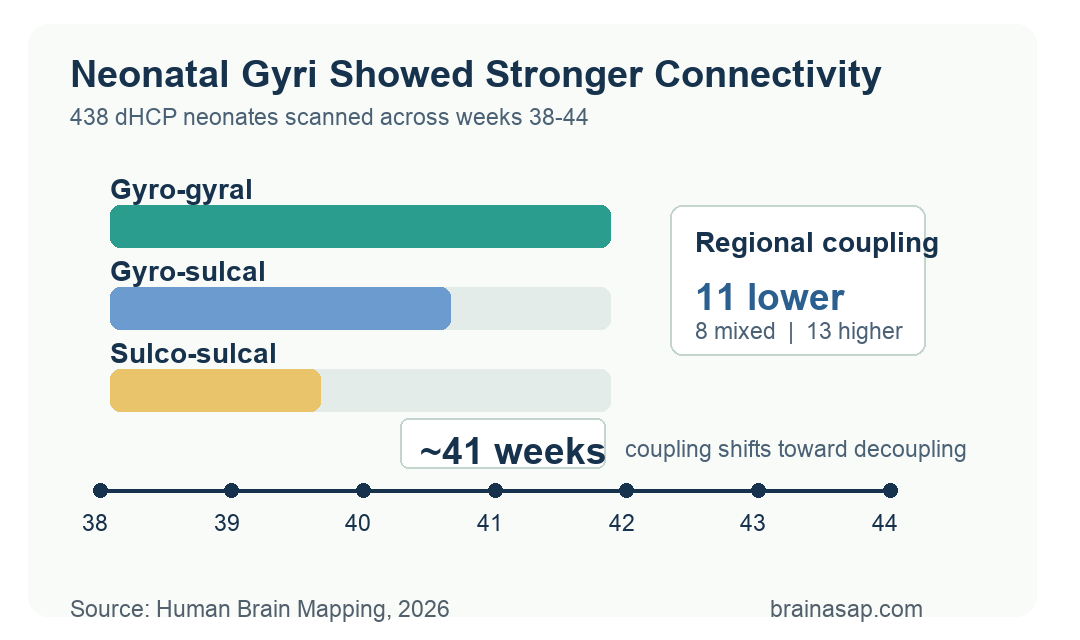
- Gyro-gyral strongest: Functional connectivity and structural connectivity were strongest between gyral regions, intermediate between gyri and sulci, and weakest between sulcal regions across the whole brain.
- 38-to-44-week pattern: The gyro-gyral > gyro-sulcal > sulco-sulcal hierarchy persisted across most lobes during the 38-to-44-week scan-age window.
- 41-week shift: Whole-brain coupling between functional and structural connectivity moved from coupled development toward decoupling after roughly 41 to 42 weeks.
Source: Human Brain Mapping (2026) | Mao et al.
Gyri and sulci are the folds and grooves that organize the cerebral cortex. The new analysis asked whether those anatomical divisions already differ in newborn brain connectivity, before months or years of postnatal experience have shaped mature brain networks.
The researchers combined structural T2-weighted MRI, diffusion-weighted MRI, and resting-state functional MRI from the developing Human Connectome Project. The work maps normative imaging patterns rather than predicting clinical outcomes for individual babies.
438 dHCP Neonates Were Scanned From 38 to 44 Weeks
The cohort included 438 full-term neonates born after 38 weeks of gestation. Scan age ranged from 38.14 to 44.71 weeks postmenstrual age, letting the team examine rapid maturation around the usual term-birth period.
Researchers used the developing Human Connectome Project because it provides harmonized multimodal neonatal imaging.
The analysis compared wiring-like structural connectivity with activity-correlation functional connectivity.
- Structural MRI: T2-weighted images defined cortical anatomy and folding.
- Diffusion MRI: Fiber reconstruction estimated structural connectivity between cortical regions.
- Resting-state fMRI: Blood-oxygen-level-dependent signal correlations estimated functional connectivity.
The cortical map started with 32 neonatal brain regions. Each region was split into a gyral part and a sulcal part, yielding 64 regions of interest for the connection matrices.
Gyro-Gyral Connectivity Was Strongest in Newborn Cortex
The main whole-brain hierarchy was consistent: gyro-gyral connections were strongest, gyro-sulcal connections were intermediate, and sulco-sulcal connections were weakest. Both functional connectivity and structural connectivity followed that order.
Gyri are often described as global information-processing hubs, while sulci are more locally specialized.
Mao et al. found that this organization was already visible in the neonatal cortex.
- Gyro-gyral: Connections between gyral regions showed the strongest mean connectivity.
- Gyro-sulcal: Mixed fold-groove connections generally sat between the two extremes.
- Sulco-sulcal: Connections between sulcal regions were weakest across the whole-brain analysis.
Lobar analyses mostly followed the same structure. Temporal, frontal, and parietal lobes showed the hierarchy clearly, while occipital and limbic results had more regional exceptions.
Functional and Structural Connectivity Changed at Different Speeds
The study also modeled how connectivity changed with scan age. Structural connectivity increased more steadily across the 38-to-44-week window, while functional connectivity followed a more curved trajectory, rising and then declining in several regions.
To quantify that relationship, the team modeled FC-SC coupling, meaning whether functional connectivity and structural connectivity were changing in the same direction over time. Positive values indicated coupled development; negative values indicated decoupling.
- Early coupling: Whole-brain FC and SC were coupled during the initial 38-to-42-week phase.
- Later decoupling: After about 42 weeks, functional and structural trajectories moved in opposing directions.
- Regional timing: The authors described a transition around 41 weeks in multiple brain regions.
The finding fits a developmental view of the newborn brain. Axonal structure can keep maturing while functional activity patterns reorganize, making the two measures temporarily diverge.
Coupling Rates Differed Across Cortical Regions
The regional analysis showed that coupling was not uniform across the cortex. Of the 32 cortical regions, 11 had coupling rates below 0.45, 8 were near balanced, and 13 had coupling rates at or above 0.55.
Those groups suggest that some regions spend more of the term-age window with structural and functional measures moving together, while others spend more time decoupled.
- Lower coupling: 11 regions had coupling less than decoupling.
- Mixed coupling: 8 regions were near a balance between the two states.
- Higher coupling: 13 regions had coupling greater than decoupling.
The occipital and limbic lobes stood out. The occipital lobe showed relatively sustained coupling, while the limbic lobe had a more distinct developmental trajectory than temporal, frontal, and parietal regions.
Neonatal Connectivity Is a Normative Map, Not a Diagnosis
The study provides a reference point for early brain organization. Fold-based connectivity differences were already present near term age, before the infant brain reaches later network maturity.
Several limits keep the finding narrow. The regions came from an atlas-level parcellation, the analysis was observational, and the work did not link connectivity patterns to later cognitive or clinical outcomes in the same infants.
- Atlas resolution: Gyri and sulci were defined from cortical curvature and region labels, not individualized developmental anatomy in every detail.
- Cross-sectional modeling: Age-related curves were modeled across scan ages rather than repeated scans for every infant.
- No outcome prediction: The study did not test whether a baby’s coupling profile predicted neurodevelopmental diagnoses.
The developmental point is anatomical as well as functional. Newborn cortical folds are not just shape; they already carry measurable differences in structural wiring, functional synchronization, and the timing of early structure-function coupling.
Citation: DOI: 10.1002/hbm.70524. Mao et al. Structuro-Functional Differentiation and Coupling of Gyri and Sulci in the Neonatal Cortex. Human Brain Mapping. 2026;47:e70524.
Study Design: Multimodal neonatal MRI analysis using structural, diffusion, and resting-state functional imaging from the developing Human Connectome Project.
Sample Size: 438 full-term neonates scanned from 38.14 to 44.71 weeks postmenstrual age, with 64 gyral/sulcal cortical regions of interest.
Key Statistic: Functional and structural connectivity were strongest for gyro-gyral pairs and weakest for sulco-sulcal pairs, with FC-SC coupling shifting toward decoupling around 41 to 42 weeks.
Caveat: The analysis maps normative neonatal connectivity patterns and does not establish an individual diagnostic or outcome-prediction test.