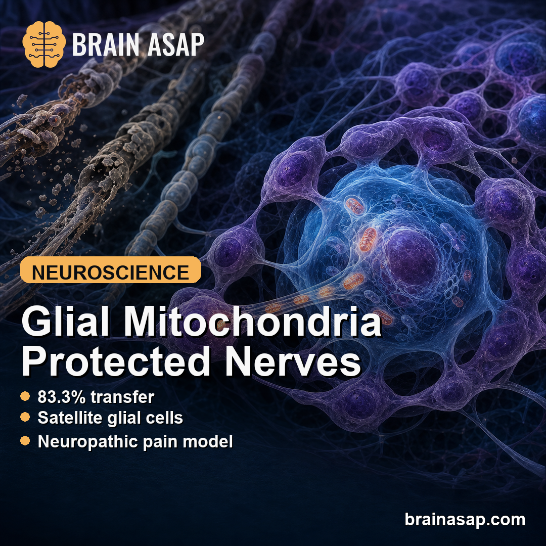
TL;DR: A 2026 Nature study found that satellite glial cells can transfer mitochondria to sensory neurons, and blocking that transfer in mice promoted nerve degeneration and neuropathic pain.
Key Findings
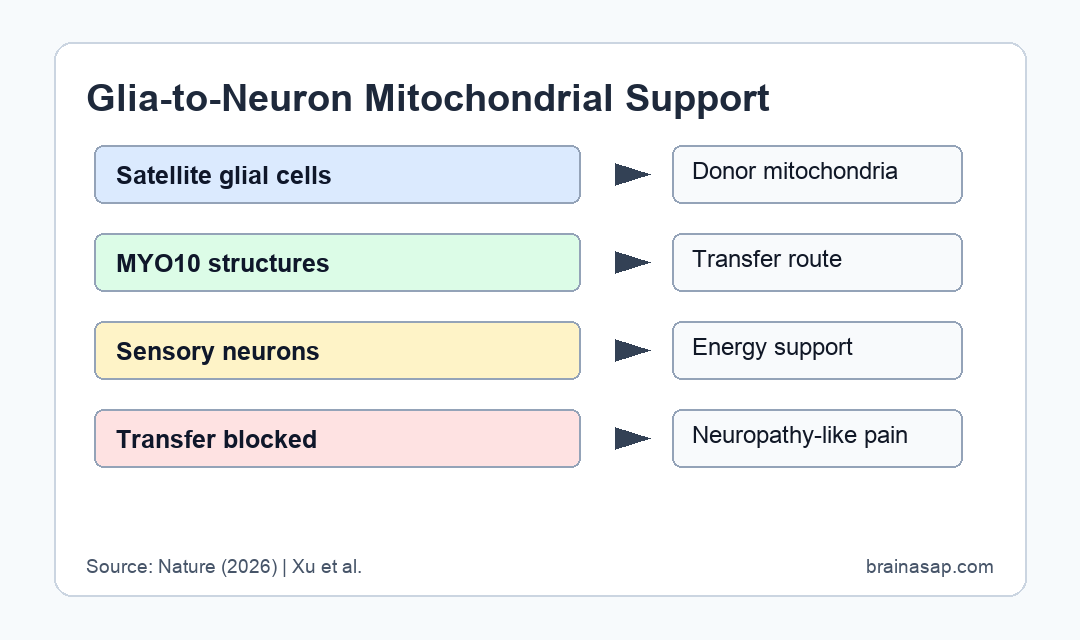
- 83.3% transfer: In mouse co-cultures, 83.3% of dorsal root ganglion neurons received mitochondria from satellite glial cells.
- 31.3% TNT-positive: Visible tunnelling nanotubes were seen in 31.3% of neurons, suggesting the structures can be transient.
- MYO10 mechanism: The transfer depended on satellite-glial myosin 10, a protein involved in forming tunnelling nanotube-like structures.
- Pain phenotype: Blocking mitochondrial transfer in naive mice led to nerve degeneration and neuropathic pain-like behavior.
- Human diabetes link: Satellite glial cells from people with diabetes showed reduced MYO10 expression and reduced mitochondrial transfer.
Source: Nature (2026) | Xu et al.
Satellite glial cells wrap around sensory-neuron cell bodies in dorsal root ganglia. This study argues that they do more than support neurons chemically; they can hand over mitochondria.
The finding gives peripheral neuropathy a concrete cell-to-cell mechanism. If sensory neurons cannot maintain enough functional mitochondria, pain signaling and nerve degeneration may follow.
Satellite Glial Cells Transferred Mitochondria to Sensory Neurons
The researchers first separated mouse satellite glial cells and dorsal root ganglion sensory neurons, labeled glial-cell mitochondria, and then placed the cells together. After 24 hours, mitochondrial signal appeared inside neurons that had not been directly labeled.
Quantitatively, 83.3% of neurons received mitochondria from satellite glial cells in the co-culture experiment. Visible tunnelling nanotubes, or TNTs, were detected in 31.3% of neurons.
- Source cells: Satellite glial cells were validated with established markers including FABP7, Kir4.1, AQP4, connexin 43, and glutamine synthetase.
- Recipient cells: The recipient cells were primary sensory neurons from dorsal root ganglia, a peripheral nervous system structure involved in pain and touch.
- Transfer route: Microscopy showed TNT-like bridges carrying mitochondria between glia and neurons.
The gap between mitochondrial-transfer rate and visible TNT rate makes biological sense. Live imaging showed that TNTs can form and disassemble within tens of minutes, so a fixed image may miss many transfer events.
MYO10 Helped Build the Glia-to-Neuron Transfer Route
The team then tested how the transfer happened. Pharmacological experiments pointed toward tunnelling nanotubes rather than simple endocytosis or gap-junction transfer.
The protein MYO10, short for myosin 10, became the central mechanism. MYO10 is involved in cellular protrusions, and reducing it in satellite glial cells decreased mitochondrial transfer.
- TNT blockade: Cytochalasin B and Y-27632 disrupted transfer-related cellular structures.
- Activity dependence: Tetrodotoxin reduced mitochondrial transfer without eliminating overall TNT formation.
- Human relevance: Single-nucleus RNA sequencing and in situ hybridization showed high MYO10 expression in human satellite glial cells.
This is not just a cell-culture observation. The paper also identified TNT-like ultrastructures between satellite glial cells and sensory neurons in mouse and human dorsal root ganglia.
Blocking Mitochondrial Transfer Promoted Neuropathic Pain
The strongest part of the paper is the functional test. When the researchers blocked mitochondrial transfer in otherwise naive mice, the animals developed nerve degeneration and neuropathic pain-like behavior.
The result suggests mitochondrial transfer is more than a rescue response after damage. Sensory neurons may need ongoing glial mitochondrial support to maintain axon health.
- Normal state: Long sensory axons have high mitochondrial demand because they must support electrical transmission over large distances.
- Blocked support: Reducing glia-to-neuron transfer impaired neuronal maintenance.
- Pain outcome: The mouse phenotype included neuropathic pain-like behavior, linking the cell mechanism to sensory symptoms.
- Therapeutic logic: Preserving or restoring transfer could be a future strategy for small-fiber neuropathy, but that remains preclinical.
The study also tested adoptive transfer of human satellite glial cells into mouse dorsal root ganglia. Those cells provided MYO10-dependent protection against peripheral neuropathy in the model.
Diabetes Reduced Human Glial Mitochondrial Transfer
Peripheral neuropathy is common in diabetes, and the paper connected the mechanism to that clinical context. Satellite glial cells from dorsal root ganglia of people with diabetes showed reduced MYO10 expression and reduced mitochondrial transfer to neurons.
The finding does not prove that this single mechanism causes diabetic neuropathy in people. It does, however, give researchers a testable cell-biological explanation for why sensory neurons may lose support in diabetes.
- Human tissue readout: The diabetes-linked reduction was observed in human dorsal root ganglion material.
- Mouse intervention: Human satellite glial cells could protect mouse sensory neurons in a MYO10-dependent way.
- Main limitation: Human clinical benefit was not tested; the therapeutic claims remain mechanistic.
The conclusion changes the cell model. Peripheral glia may act as active mitochondrial donors, not passive bystanders, in the maintenance of sensory-neuron health.
Long Sensory Axons Make Mitochondrial Support Especially Important
Dorsal root ganglion neurons have long peripheral branches, and human sensory axons can extend across large distances. That geometry creates a practical energy problem: mitochondria must support terminals far from the cell body.
The study’s glia-to-neuron transfer mechanism fits that anatomy. If a sensory neuron cannot generate, transport, or maintain enough healthy mitochondria, a nearby satellite glial cell may provide support where demand is high.
- Peripheral-neuron burden: Long axons need reliable energy supply for signaling, repair, and terminal maintenance.
- Glial proximity: Satellite glial cells closely surround sensory-neuron cell bodies, putting them in position to exchange material.
- Disease clue: Diabetes-linked reduction in MYO10 and transfer offers a plausible route from metabolic disease to sensory-neuron vulnerability.
That framework also helps explain why a local support-cell defect could produce widespread symptoms. Pain fibers and small sensory fibers are metabolically demanding, and a failure in mitochondrial support may show up as neuropathic pain before gross nerve loss is obvious.
Citation: DOI: 10.1038/s41586-025-09896-x. Xu et al. Mitochondrial transfer from glia to neurons protects against peripheral neuropathy. Nature. 2026;650:951-960.
Study Design: Cell-culture, mouse-model, microscopy, human-tissue, and adoptive-transfer study.
Sample/Model: Mouse and human dorsal root ganglia, satellite glial cells, sensory neurons, and neuropathy models.
Key Statistic: In co-culture, 83.3% of sensory neurons received glial mitochondria, while 31.3% showed visible TNTs.
Caveat: The work identifies a mechanism and preclinical protection, not a tested treatment for human neuropathy.