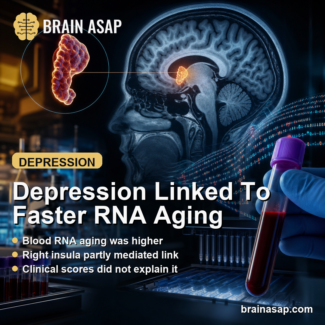
TL;DR: A 2026 case-control study in Psychological Medicine linked major depressive disorder to faster blood-based transcriptomic aging and found that right-insula brain changes partly explained the depression association.
Key Findings
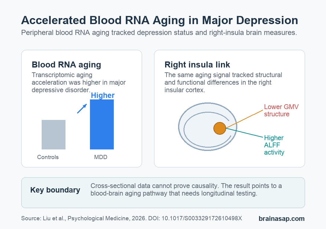
- Transcriptomic aging was faster in depression: 141 people with major depressive disorder showed higher blood RNA-based aging acceleration than 134 healthy controls.
- The result strengthened after adjustment: the depression group difference was significant before adjustment and stronger after accounting for chronological age and sex.
- Right-insula measures tracked the aging marker: transcriptomic aging was associated with lower right-insula gray matter volume and higher right-insula resting-state activity.
- Clinical severity did not explain it: transcriptomic aging was not significantly linked to depression symptoms, anxiety symptoms, cognition, childhood trauma, or polygenic risk score.
- The design was cross-sectional: the study points to a blood-brain aging pathway in depression but cannot prove that transcriptomic aging causes depression.
A blood-based aging marker looked older in people with major depressive disorder. The brain finding that connected most clearly to that marker was not a broad whole-brain effect.
It was a right-insula pattern involving both structure and resting-state function.
The study used peripheral blood RNA sequencing to estimate transcriptomic age, meaning biological age inferred from gene-expression patterns rather than from a birth date. Researchers then compared that estimate with chronological age to calculate transcriptomic aging acceleration.
Blood RNA Suggested Faster Biological Aging in Depression
The sample included 141 people with major depressive disorder (MDD) and 134 healthy controls. Major depressive disorder is a clinical depression diagnosis; in this study, participants were Han Chinese, ages 16-40, and were either antidepressant-naive or had stopped antidepressants for at least 3 months.
Researchers built a transcriptomic-age model from genes associated with chronological age, then asked whether the predicted RNA-based age deviated from actual age. That deviation was higher in the MDD group.
- Before adjustment: transcriptomic aging acceleration was higher in MDD than controls (t = 2.06, P = 0.040).
- After age and sex adjustment: the difference was stronger (t = 3.72, P < 0.001).
- Interpretation: the depression group looked biologically older by this blood RNA measure, even though the study was not designed to prove cause and effect.
Depression has often been discussed alongside inflammation, mitochondrial stress, cognitive aging, and age-related disease risk. Researchers examined those links with a blood gene-expression estimate of biological aging rather than symptom ratings alone.
The authors did not frame the RNA clock as a diagnostic test for depression. It is better read as a biological aging marker that may help describe one pathway of vulnerability.
Inflammation and Energy Pathways Overlapped With Aging
The enrichment analysis compared biological processes tied to age-associated genes and MDD-associated genes. Several overlapping pathways pointed toward systems already implicated in both aging and depression.
- Innate immune inflammation: aging-associated and MDD-associated transcriptomic patterns both involved innate immune-related processes.
- Ribosome biogenesis: the study found overlap in machinery related to ribosome production, which can be disrupted during cellular stress.
- Mitochondrial energy metabolism: oxidative phosphorylation and mitochondrial gene-expression pathways appeared in both aging and MDD analyses.
- Telomere maintenance: telomere-related enrichment appeared more specific to aging than to MDD in this dataset.
The inflammation result is the most reader-facing part of the pathway analysis. The authors describe innate immune-related inflammation as a possible bridge between aging biology and depression biology, not as a standalone explanation for every case of MDD.
The telomere finding narrows the claim. The study did not show that every hallmark of aging maps neatly onto depression.
Some transcriptomic aging processes overlapped with MDD, while telomere maintenance looked more aging-specific in this analysis.
Right-Insula Structure and Activity Carried the Brain Link
The researchers also analyzed MRI measures. Gray matter volume (GMV) measured structural tissue volume, while amplitude of low-frequency fluctuations (ALFF) measured resting-state brain activity.
Transcriptomic aging was associated with right-insula GMV (t = -3.30, P = 0.001) and right-insula ALFF (t = 2.64, P = 0.009). In simpler terms, the blood aging readout tracked both lower structure and higher resting-state functional activity in the right insular cortex.
The insula is involved in interoception, emotional processing, salience, and the integration of internal body states. That role makes it a plausible region to test when a peripheral biological marker is being connected with depression vulnerability.
The mediation result was narrower than a headline might suggest. Right-insula abnormalities partially mediated the association between transcriptomic aging and MDD.
Partial mediation means the insula explained some of the statistical relationship, not all of it.
Symptoms, Cognition, Trauma, and Genetic Risk Did Not Explain the Aging Readout
One of the more important negative findings was that transcriptomic aging did not significantly track several familiar depression measures. That included clinical symptom severity, neurocognitive performance, childhood trauma scores, and MDD polygenic risk score.
Those null results make the interpretation more specific. The RNA-aging marker was not simply a proxy for worse current depression scores, higher anxiety scores, poorer cognition, reported childhood trauma, or inherited common-variant risk as measured here.
- Clinical symptoms: depression and anxiety ratings did not show significant associations with transcriptomic aging.
- Cognition: Cambridge Neuropsychological Test Automated Battery measures did not explain the RNA-aging association.
- Risk history: childhood trauma score and polygenic risk score were not significantly associated with transcriptomic aging.
The authors suggest that transcriptomic aging may be a more integrated marker shaped by long-term gene-environment biology rather than a short-term episode marker.
That interpretation remains a hypothesis until longitudinal studies test whether the RNA-aging readout changes before, during, or after depressive episodes.
The Case-Control Design Limits the Claim
The study was carefully layered, but its design still sets real boundaries. It was a single-center case-control study with no longitudinal follow-up, so it cannot show whether transcriptomic aging came before depression, followed depression, or developed alongside it.
Several other caveats matter for interpretation:
- Sample scope: all participants were recruited from one center, and generalizability to other populations needs replication.
- Model construction: gene selection and model construction were done within the same sample, which can make age prediction look stronger than it would in an outside dataset.
- Missing covariates: smoking status and body mass index were not included, even though both can affect peripheral gene expression.
- Transcriptome scope: the analysis focused on mRNA expression and did not include microRNAs, long noncoding RNAs, or other transcriptomic layers.
The most defensible takeaway is that depression was associated with faster blood RNA-based aging and with right-insula brain differences that partly linked that aging marker to MDD status.
The next question is whether this marker predicts future depression risk, treatment response, relapse, or biological recovery over time.
Citation: DOI: 10.1017/S003329172610498X. Liu et al. Peripheral transcriptomic aging acceleration in major depressive disorder: the mediating role of insular cortex alterations. Psychological Medicine. 2026;56.
Study Design: Single-center case-control study using peripheral blood RNA sequencing, clinical measures, genetic risk scoring, and MRI analyses.
Sample Size: 141 people with major depressive disorder and 134 healthy controls.
Key Statistic: Transcriptomic aging acceleration was higher in MDD after adjustment for chronological age and sex (t = 3.72, P < 0.001).
Caveat: The study was cross-sectional and cannot prove that transcriptomic aging causes depression.