How Gene Therapy Mimicked Morphine for Chronic Pain
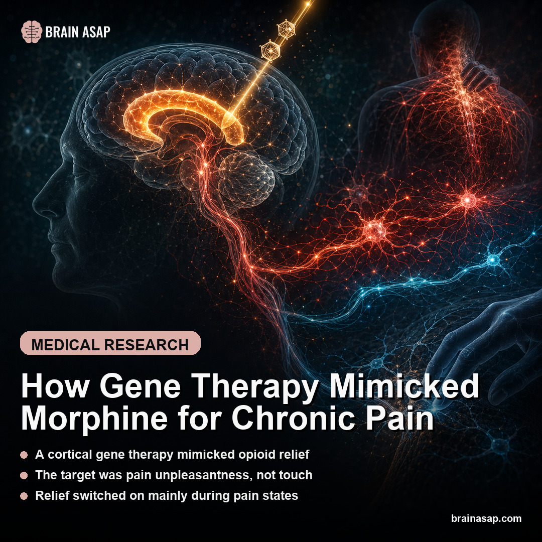
TL;DR: A Nature study identified opioid-sensitive neurons in the anterior cingulate cortex and used a synthetic mu-opioid receptor promoter to silence them, producing morphine-like relief of chronic pain unpleasantness in mice without using a systemic opioid drug.
Key Findings
- 700-μm nociceptive hotspot: The authors mapped a narrow pain-responsive zone in the anterior cingulate cortex and found that the key glutamatergic clusters expressing Oprm1 stayed opioid-accessible even after chronic nerve injury.
- ACC opioid receptors were decisive: Knocking out mu-opioid receptors specifically in the anterior cingulate erased morphine’s affective analgesia, while re-expressing them there restored it, showing this cortical circuit was both necessary and sufficient for that part of pain relief.
- 20 body points became a pain scale: Using a deep-learning system called LUPE, the team tracked 20 mouse body points, classified 6 spontaneous behaviors, and distilled them into an affective-motivational pain scale that rose with injury and fell with morphine.
- 5-mouse calcium imaging linked neurons to pain state: Single-cell recordings showed that anterior cingulate neurons tracking spontaneous pain-related licking were selectively reshaped by morphine during acute and chronic pain states.
- MORp gene therapy matched morphine: A synthetic mu-opioid receptor promoter driving inhibitory chemogenetics reproduced morphine-like reductions in pain states during neuropathic pain and, in some heat and cold assays, outperformed systemic 0.5 mg/kg morphine.
- Relief without rewarding healthy mice: Inhibiting these neurons was reinforcing for mice with nerve injury, which is what you would expect from pain relief, but did not create place preference in uninjured mice.
Source: Nature (2026) | Oswell et al.
Opioids work, but they work messily. They dull the suffering component of pain while also touching breathing, reward, constipation, tolerance, and addiction risk. This paper asked a more surgical question: what if you could isolate the exact cortical pain circuit opioids calm down, then turn off only that circuit on demand?
The Anterior Cingulate Matters More for Pain Unpleasantness Than Pain Detection
One of the most important ideas in pain neuroscience is that pain intensity and pain unpleasantness are not the same thing. You can feel a stimulus and still suffer less from it, which is exactly why cingulotomy once helped some patients with intractable pain without making them numb to every harmful sensation.
This paper builds directly on that idea. The target was the anterior cingulate cortex, especially a pain-responsive hotspot where the authors found neurons expressing the mu-opioid receptor, the same receptor morphine binds. In other words, the team was not looking for a vague “pain area.” They were looking for the cortical ensemble most plausibly responsible for the part of pain opioids actually soften.
The reason is modern analgesic development has spent decades chasing molecules more than circuits. If the emotionally aversive part of chronic pain lives in a relatively defined cortical network, then a therapy aimed at that network could, at least in theory, keep the benefit while shedding some of the systemic baggage of opioids.
How a 20-Body-Point Tracking System Turned Mouse Behavior Into an Affective Pain Score
To get there, the authors first had to solve a measurement problem. Standard rodent pain assays lean heavily on reflexes such as paw withdrawal.
Useful, yes, but crude. They tell you whether an animal reacts, not whether the animal is trapped in an ongoing aversive state that feels more like chronic pain in humans.
The team built a platform called LUPE, which tracked 20 body points in freely moving mice, classified six broad behaviors, and then used transition patterns among those behaviors to infer latent pain states. In acute formalin and capsaicin experiments, each condition included 20 mice, and the system extracted a principal-component measure that the authors called the affective-motivational pain scale, or AMPS.
That distinction is the paper’s first real innovation. Morphine did not just flatten everything.
It selectively suppressed the behavioral dimension that rose with injury and tracked affective pain. The authors argue this gives them something richer than a reflex metric: a readout of how much the animal is inhabiting a pain state rather than merely noticing a noxious event.
For a field that often overpromises translation from simple animal assays, the measurement upgrade is important almost as much as the therapy itself. If your pain scale cannot distinguish sensory detection from pain burden, you cannot really claim to have built a safer analgesic.
How 0.5 mg/kg Morphine Exposed a Mu-Opioid Circuit in the Cingulate
The most convincing causal experiment in the paper was not the gene therapy. It was the receptor logic.
When the authors deleted mu-opioid receptors specifically from anterior cingulate neurons, a low morphine dose of 0.5 mg/kg no longer reduced affective-motivational pain responses. Then they reversed the experiment: re-expressed those receptors in the same cortical region of global knockout mice and brought the analgesic effect back.
The sequence is important because it narrows the mechanism dramatically. The paper is not just saying the anterior cingulate lights up during pain or quiets down after opioids. It shows that morphine’s relief of pain unpleasantness depends on opioid receptor action inside that cortical ensemble.
Single-nucleus RNA sequencing added another layer. In the nociceptive hotspot, the authors resolved 23 cell types, but only three glutamatergic clusters showed persistent nociceptive and neuropathic immediate-early-gene signatures, and all three expressed Oprm1. That is a satisfyingly specific result: not all cingulate cells matter equally, and the ones that look most pain-relevant also look opioid-relevant.
The implication is subtle but important. Opioids may work less by muting raw sensory input than by quieting a cortical network that tags pain as urgent, aversive, and behaviorally compelling. That is exactly the component drug developers would love to separate from respiratory depression and reward effects.
What 5-Mouse Calcium Imaging Revealed About Pain-Tracking ACC Neurons
The paper then moved from cell identity to live circuit dynamics. In 5 mice carrying calcium indicators and implanted GRIN lenses, the authors recorded anterior cingulate activity while the animals experienced acute capsaicin pain and then morphine analgesia inside the LUPE chamber.
They identified neurons whose activity predicted the probability of pain-related licking, a motivated behavior the authors interpret through the gate-control framework as part of an attempt to regulate pain. These Plick neurons were not random bystanders. Their activity changed with injury, changed with morphine, and predicted how much time the animal spent in pain-linked states.
That is one reason the paper feels stronger than a typical circuit claim. It did not stop at “this neuron type exists.” It showed that spontaneous behavior, latent pain-state occupancy, and neuronal activity co-moved across acute and chronic pain. By 3 weeks after spared nerve injury, those lick-linked neural responses were blunted, and morphine partially restored them.
In other words, chronic pain in this model was not simply more firing. It was a reorganization of the behavioral-neural relationship inside the anterior cingulate. Morphine appeared to rescue that relationship, which fits the paper’s core claim that opioid analgesia is changing the affective computation of pain rather than merely numbing input.
How a Synthetic Mu-Opioid Promoter Became a Gene Therapy for Chronic Pain
Once the authors knew which cortical ensemble morphine was calming, they tried to mimic the same effect without giving morphine. Their tool was a synthetic mu-opioid receptor promoter, or MORp, designed to drive expression selectively in opioid-sensitive anterior cingulate neurons. Into those cells they delivered an inhibitory chemogenetic construct, hM4Di, which could later be activated with deschloroclozapine.
The logic is elegant. Instead of delivering an opioid systemically and hoping the right cells are hit among many wrong ones, deliver a genetic actuator only to the right cells, then turn those cells down on command. In neuropathic pain experiments, this MORp-hM4Di approach changed three pain-state readouts:
- Injured-paw licking: mice spent less time showing spontaneous pain-linked behavior.
- Pain-state occupancy: LUPE classified less time in latent pain states.
- AMPS score: the affective-motivational pain scale moved downward.
The comparison against morphine is where the paper gets ambitious. In the key assays, the gene-therapy strategy matched morphine on affective pain relief and even beat it in some injured-mouse tests involving noxious heat and cold. It did not change mechanical von Frey thresholds, which is consistent with the larger thesis: the intervention is best at the unpleasant, motivational dimension of pain, not a universal sensory shutdown.
The authors also showed that chemogenetic inhibition reduced brain-wide neuropathic activity downstream of anterior cingulate projections. So the therapy did not just change one local signal. It appeared to calm a broader pain network by acting at one strategically important cortical entry point.
State-Dependent Relief Matters More Than a Mouse Pain Headline
The addiction question hangs over every opioid-adjacent paper, so the most reassuring result may be what happened in the place-preference assay. Silencing these anterior cingulate MOR-positive neurons was reinforcing in mice with nerve injury, which is what you would expect if the intervention relieved ongoing suffering. But the same manipulation did not create place preference in uninjured mice.
That is not the same as proving the approach is addiction-proof. It is a mouse assay, not a human safety dossier.
Still, the state dependence matters. A treatment that feels rewarding only when pain is present is fundamentally different from a drug that becomes rewarding in healthy tissue too.
The remaining translational leap is enormous. This is a preclinical mouse study using viral tools, invasive access, and synthetic promoter engineering. The authors point toward future noninvasive delivery ideas, including focused ultrasound blood-brain barrier opening, but that is clearly future-facing speculation, not a next-year clinic plan.
So the right way to read the paper is not “gene therapy cures chronic pain.” It is narrower and more useful: opioid analgesia can be reverse-engineered at the circuit level. If the suffering component of chronic pain depends heavily on a defined anterior cingulate ensemble, then safer analgesics may come from targeting that ensemble directly rather than flooding the whole body with opioid molecules.
Paper: Mimicking opioid analgesia in cortical pain circuits. Nature. 2026;649:938-946.. DOI: 10.1038/s41586-025-09908-w
Authors: Oswell et al.
Study Design: Preclinical animal study
Sample Size: 700-μm nociceptive hotspot: The authors mapped a narrow pain-responsive zone in the anterior cingulate cortex and found that the key glutamatergic clusters expressing Oprm1 stayed opioid-accessible even after chronic nerve injury.
Key Statistic: ACC opioid receptors were decisive: Knocking out mu-opioid receptors specifically in the anterior cingulate erased morphine’s affective analgesia, while re-expressing them there restored it, showing this cortical circuit was both necessary and sufficient for that part of pain relief.