How Astrocytes Help the Amygdala Store Fear Memories
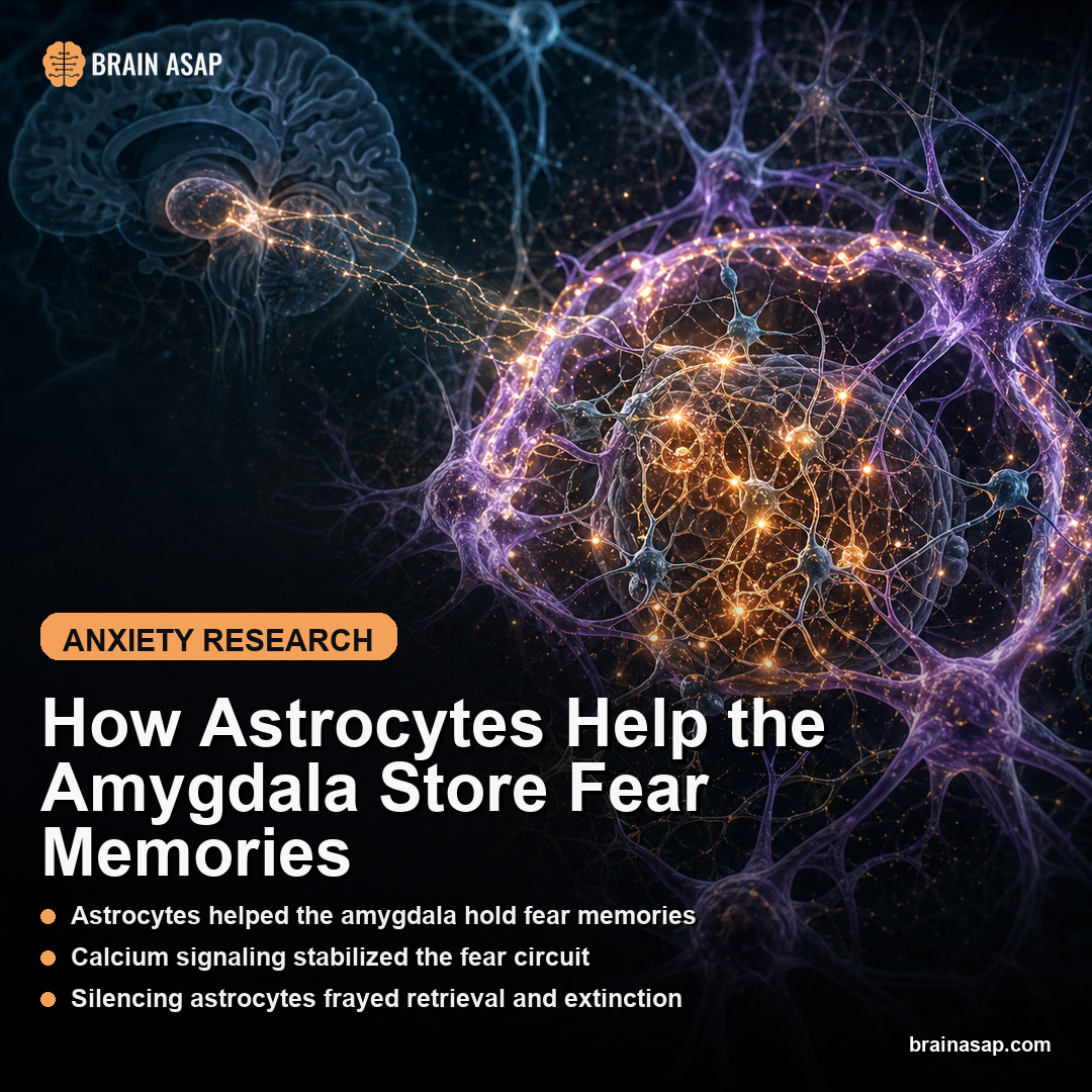
TL;DR: Amygdala astrocytes were not passive support cells: their calcium signals tracked learned fear states and were required for neuronal fear-memory representations in mice.
Key Findings
- In vivo BLA imaging across multiple mouse cohorts: The team combined astrocyte and neuronal calcium imaging, electrophysiology, and circuit manipulations in the basolateral amygdala to test whether glia participate in fear-memory coding rather than just supporting it metabolically.
- Astrocyte activity tracked the fear state itself: During conditioned-fear retrieval and extinction, BLA astrocytes showed dynamic calcium patterns that shifted with the animal’s behavioral state instead of remaining a slow background signal.
- Retrieval and extinction both depended on astrocyte calcium: When astrocyte signaling was perturbed, the neuronal population patterns that normally encode fear retrieval and extinction were degraded, and behavior changed with them.
- BLA-to-prefrontal readout also broke down: The paper tied astrocyte signaling to a BLA-prefrontal circuit, suggesting the glial contribution is not local decoration but part of how memory-related output reaches downstream decision-making regions.
- The paper revises a neurocentric model: Instead of treating astrocytes as passive support cells, the study argues they help generate and update the neural representations that underlie adaptive threat responses.
Source: Nature (2026) | Bukalo et al.
Fear circuits are usually told as a neuron-only model. This paper makes that version feel incomplete. The cells doing the housekeeping around amygdala synapses turned out to be part of the code.
Why Fear-Memory Models Kept Missing the Cells Wrapped Around the Synapse
The modern fear-memory model usually begins in the basolateral amygdala. A cue that once predicted danger comes back, a neuronal ensemble reactivates, and the animal freezes.
Later, after extinction training, a partially different ensemble helps encode the updated “this cue is now safe” meaning. It is a powerful model, but it is also a neuron-heavy one.
That bias is exactly what makes this paper interesting. Astrocytes sit on top of the same synapses, sense neurotransmitters, regulate glutamate clearance, and run their own calcium dynamics.
Yet in most psychiatric circuit models they are still treated like infrastructure rather than like participants. Bukalo and colleagues asked a more direct question: what if the cells hugging the synapse are part of the representation itself?
The answer is not subtle. In this study, astrocyte activity in the amygdala did not just correlate with the animal being in a fear experiment.
It tracked whether fear was being retrieved, extinguished, or updated. And when the investigators pushed on astrocyte calcium signaling directly, the underlying neuronal map became harder to sustain.
What Amygdala Imaging Measured During Fear Retrieval
This was a dense mechanistic paper, but the design idea was straightforward. The authors combined in vivo calcium imaging, electrophysiology, and causal manipulations of astrocytes in mice undergoing conditioned-fear retrieval and extinction. That let them compare three layers at once: what the animal was doing, what astrocytes were doing, and what nearby neurons were doing.
The abstract-level result is the cleanest summary: BLA astrocytes dynamically tracked fear state and supported both fear-memory retrieval and extinction. In other words, these glial cells were not only active during emotionally salient moments. Their activity changed in ways that mapped onto the same memory operations neuroscientists usually assign to neurons alone.
The reason is the amygdala is not simply flipping between “on” and “off.” Retrieval and extinction are distinct computations. Retrieval pulls the old threat meaning back online.
Extinction gradually builds a competing safety representation. A cell type that helps sustain both sides of that transition is influencing the circuit at a high level, not just tuning background excitability.
The paper also matters methodologically. It did not stop at calcium movies.
The authors layered in electrophysiological recordings and circuit-level readout, which makes the interpretation less vulnerable to the classic criticism that glial signals are epiphenomenal and too slow to matter. Their argument is that astrocyte calcium participates in the transformation of memory state into a population code the circuit can actually use.
- Astrocyte imaging: tracked glial calcium activity as fear memories were retrieved or extinguished.
- Neuronal readout: tested whether nearby fear-memory ensembles stayed organized when astrocyte signaling changed.
- Circuit output: linked local amygdala changes to downstream prefrontal communication.
How Silencing Astrocyte Calcium Frayed Fear Retrieval and Extinction
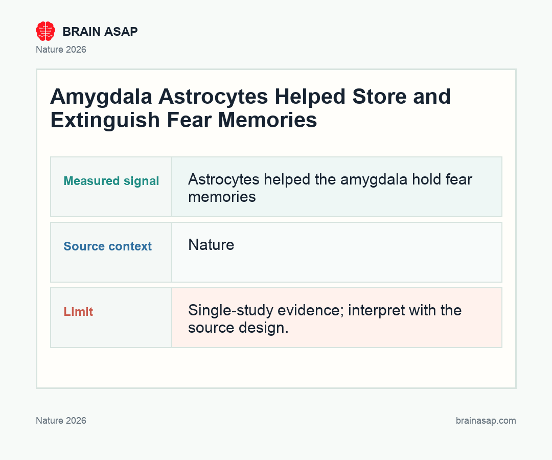
The most persuasive part of the paper is the causal step. Once the team manipulated astrocytes, the neuronal representations that normally carry retrieval and extinction information no longer looked intact. That is the pivot from “astrocytes are active” to “astrocytes are necessary.”
Conceptually, the result says the neuronal ensemble is not a self-sufficient object. It depends on the chemical and synaptic context created by nearby astrocytes.
The biology fits the result. Astrocytes regulate glutamate spillover, potassium buffering, and gliotransmitter release, all of which can reshape how a local network stabilizes a particular firing pattern.
The striking implication is that a fear memory does not necessarily live entirely inside a stable set of neurons. It may live partly inside a neuron-glia partnership that keeps the relevant ensemble coherent at the moment the cue returns. That is a more distributed and less romantic model of memory than the classic “engram cell” model, but it is probably closer to how real tissue works.
For extinction, the point may be even more important. Extinction is not deleting the old memory.
It is building a new pattern that competes with it. If astrocytes help the circuit switch between those states, they become plausible players in disorders where fear updating goes wrong, including PTSD and chronic anxiety disorders.
The BLA-to-Prefrontal Handoff Matters More Than a Local Amygdala Effect
The paper did not end at the amygdala. By linking astrocyte manipulations to readout through a BLA-prefrontal circuit, the authors showed that the glial effect is important for downstream communication, not only for local microcircuit function.
The distinction is important for psychiatry. A lot of symptoms are less about whether a brain region activates in isolation and more about whether one region can update another.
The prefrontal cortex is central to regulation, context, and behavioral flexibility. If the amygdala cannot send a properly updated signal downstream, extinction-like learning may fail at the network level even if individual neurons still spike.
The study therefore invites a richer causal chain. Traumatic learning is stored and retrieved through neuronal populations, but the stability and flexibility of those populations may depend on astrocytic calcium dynamics. That, in turn, shapes whether the amygdala can tell prefrontal cortex, “the danger memory is active” or “the safety update now deserves priority.”
It is also the kind of result that helps explain why some interventions that look powerful at the receptor level do not translate neatly into durable behavioral change. If memory updating depends on a multicellular circuit state, not just on neuronal firing, the target space for future treatments gets wider.
How a Mouse Circuit Paper Still Changes the Psychiatric Conversation
This is still a mouse study, and it does not deliver a near-term therapeutic protocol. No one should read it as proof that glia-targeting drugs are ready to improve human exposure therapy next year. But that is not the standard it needs to meet.
The more important contribution is conceptual. Psychiatric neuroscience has spent decades building neuron-first maps of fear, reward, and memory.
Those maps have been useful, but they also leave a lot of unexplained variance. Astrocytes are abundant, excitable in their own way, and anatomically positioned to shape the exact synapses we care about. A paper like this says we probably underfit the system when we leave them out.
There is also a translational hint here. If pathological fear persistence reflects not just a bad memory trace but a failure of adaptive representational updating, then therapies aimed at extinction could eventually be judged by whether they restore neuron-glia coordination, not just by whether they suppress overt fear behavior in the moment.
The cleanest way to say it is this: the amygdala did not stop being a neuronal circuit in this paper. It became a multicellular one. And once you see retrieval and extinction that way, old psychiatric models of fear look a little too simple.
Paper: Astrocytes enable amygdala neural representations supporting memory. Nature. 2026;652:434-442.. DOI: 10.1038/s41586-025-10068-0
Authors: Bukalo et al.
Study Design: Cohort study
Sample Size: In vivo BLA imaging across multiple mouse cohorts: The team combined astrocyte and neuronal calcium imaging, electrophysiology, and circuit manipulations in the basolateral amygdala to test whether glia participate in fear-memory coding rather than just supporting it metabolically.
Key Statistic: Astrocyte activity tracked the fear state itself: During conditioned-fear retrieval and extinction, BLA astrocytes showed dynamic calcium patterns that shifted with the animal’s behavioral state instead of remaining a slow background signal.