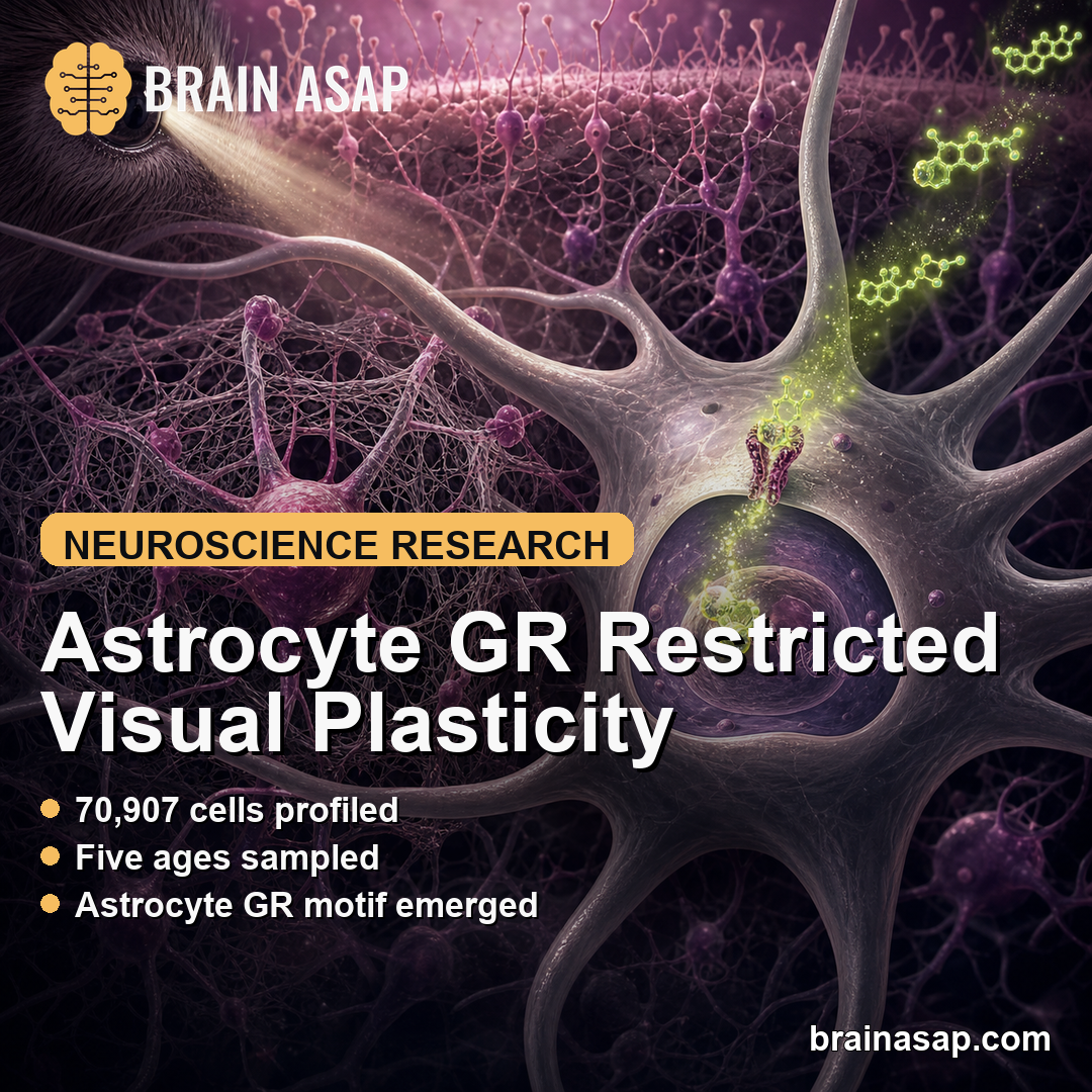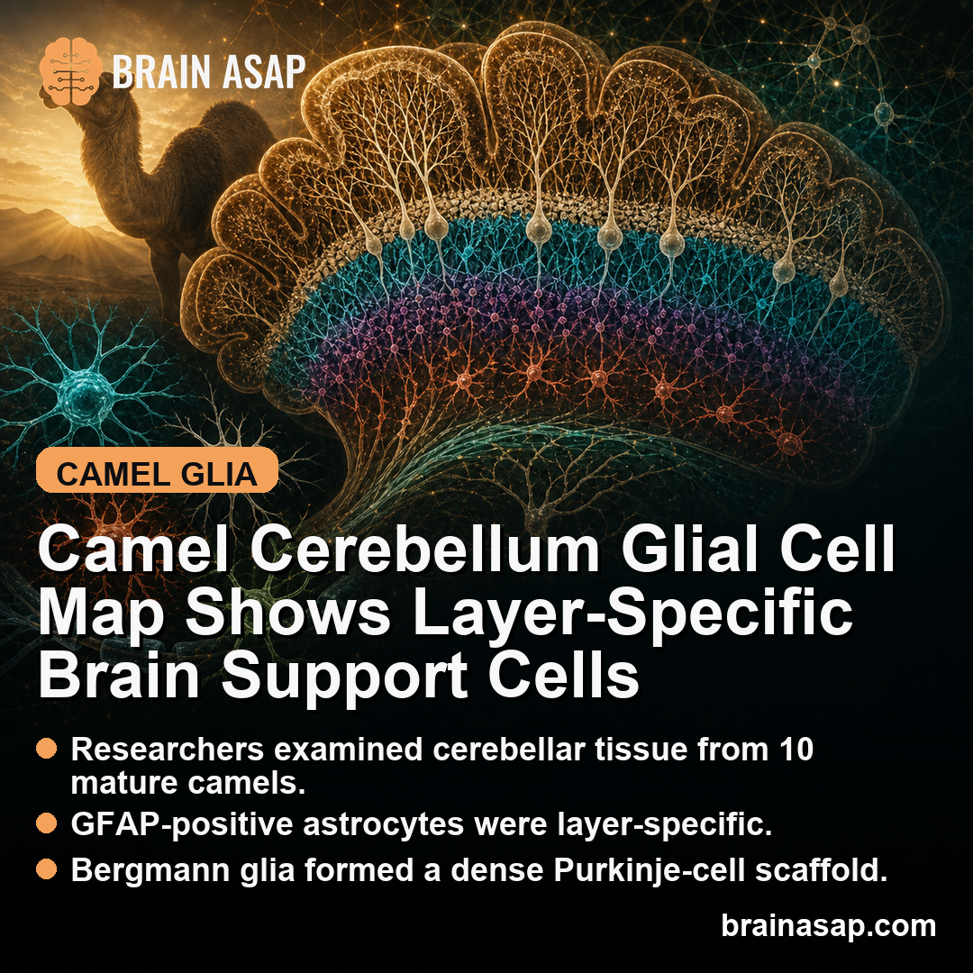
TL;DR: A 2026 Scientific Reports study mapped astrocytes, oligodendrocytes, Bergmann glia, and microglia in the camel cerebellum, showing that glial support cells are distributed very differently across cerebellar layers.
Key Findings
- 10 mature camels: Researchers examined cerebellar tissue from 10 mature male camel heads.
- GFAP was layer-specific: GFAP-positive astrocytes appeared in granular layer and white matter, but not in the molecular layer.
- White matter had more astrocyte coverage: GFAP area coverage averaged 35% in white matter versus 12% in granular layer.
- Bergmann glia formed a dense scaffold: Bergmann glia clustered in 4 to 6 rows near Purkinje cells, with density around 5,126 cells/mm2.
- Microglia varied by layer: Iba1-positive microglia were densest in white matter and granular layer and lowest in molecular and Purkinje layers.
Source: Scientific Reports (2026) | Attaai et al.
Camel Cerebellum Tissue Offered a Comparative Glia Map
The cerebellum is best known for coordination, balance, and fine motor control, but it is also involved in learning, timing, and cognitive processing.
Most public attention goes to neurons, especially Purkinje cells. This study focused instead on glia, the support-cell populations that help maintain, insulate, protect, and regulate neural circuits.
The study is not a disease trial and does not test a treatment. Its value is anatomical: it gives researchers a layer-by-layer map of glial cells in the camel cerebellar cortex.
Comparative brain anatomy can help show which cellular arrangements are conserved across mammals and which may reflect species-specific adaptations.
Camels are physiologically unusual animals, with adaptations for heat, dehydration, and long-distance movement. Many brain features will still be shared across mammals, but comparative neurobiology can test whether neural support systems differ in measurable ways.
Glial maps also create a baseline for later pathology studies. Without a normal reference pattern, it is hard to know whether a disease, injury, toxin, or aging process has changed the cerebellum.
A species-specific atlas helps future researchers separate ordinary layer organization from abnormal remodeling.
GFAP, Olig2, S-100, and Iba1 Marked Different Glial Cell Types
Researchers used histochemical and immunohistochemical staining to map several glial markers.
- GFAP: Glial fibrillary acidic protein, a marker commonly used to identify astrocytes and related glial structures.
- Olig2: A marker for oligodendrocyte-lineage cells, which are important for myelin formation and maintenance.
- S-100: A marker that highlighted Bergmann glia in the cerebellar cortex.
- Iba1: A marker for microglia, the brain’s resident immune surveillance cells.
Bergmann glia are specialized astrocyte-like cells in the cerebellum. They extend long radial processes through the molecular layer and help organize the cerebellar cortex.
The researchers examined tissue across the classic cerebellar layers: molecular layer, Purkinje layer, granular layer, and white matter. Each zone contains different cell bodies, axons, synapses, and support requirements.
Using multiple markers was important because no single stain can describe the glial environment.
GFAP, Olig2, S-100, and Iba1 point to different cellular roles, so the combined map gives a more complete picture of tissue support, myelin maintenance, radial scaffolding, and immune surveillance.
GFAP Astrocyte Coverage Was Highest in Camel Cerebellar White Matter
The GFAP result was strongly layer-specific. GFAP-positive astrocytes were absent from the molecular layer, present in the granular layer, and especially prominent in white matter.
Quantitatively, GFAP-positive area coverage averaged 35% in white matter, with a wide range from 1% to 79%. In the granular layer, coverage averaged 12% , with a range from 4% to 26%.
The layer difference was statistically significant, with F(1,20) = 8.22 and p = 0.0095. Higher GFAP coverage in white matter fits the different jobs of those regions.
White matter contains many myelinated axons and requires intense metabolic and structural support.
Granular layer contains dense neuronal cell bodies and local circuitry, while molecular layer is dominated by synaptic architecture and long processes.
The absence of GFAP signal in the molecular layer should not be read as an absence of glial influence. Bergmann glial fibers extend through that layer, and other glial markers can capture support-cell architecture that GFAP alone does not show.
The key point is that astrocyte-marker expression differed sharply by compartment.
Bergmann Glia Formed a Dense Purkinje-Layer Scaffold
Bergmann glia were one of the most visually organized findings. Researchers described them as arranged in 4 to 6 rows in the Purkinje cell layer, with processes extending through the molecular layer toward the pial surface.
The reported Bergmann glia density was about 5,126 cells/mm2. Their radial processes averaged 323 +/- 28 micrometers in length, spanning the molecular layer.
Under the microscope, neurons, glia, and processes occupied repeating layered positions.
Functionally, Bergmann glia help regulate extracellular ions and neurotransmitters, guide developmental organization, and support Purkinje cell function. The camel findings do not prove a special behavioral role, but they confirm that this highly structured glial scaffold is clear in camel cerebellar cortex.
This is especially relevant because Purkinje cells are the main output neurons of the cerebellar cortex. A dense Bergmann glial arrangement around them suggests a carefully organized support environment for cells that integrate large amounts of synaptic input.
Oligodendrocytes and Microglia Followed Distinct Layer Patterns
Olig2-positive oligodendrocytes appeared mainly in white matter and granular layer. That distribution is expected because oligodendrocytes support myelinated axons, and white matter is rich in long fiber tracts.
Iba1-positive microglia also varied by layer. Across the cerebellar cortex, mean microglia density was 178 +/- 57 cells/mm2.
White matter had the highest density at 205 +/- 48 cells/mm2, followed by granular layer at 179 +/- 25 cells/mm2.
Molecular layer was lower at 88 +/- 15 cells/mm2. That lower density fits a region dominated by synaptic processes rather than dense cell bodies or fiber tracts.
The layer effect was statistically strong, with F(3,17) = 12.44 and p = 0.00015.
The researchers reported higher microglia density in medullary white matter than molecular and Purkinje layers, and higher density in granular layer than molecular and Purkinje layers.
Microglia are often discussed in the context of inflammation or neurodegeneration, but that would be the wrong frame here. In healthy tissue mapping, microglia density helps describe normal immune surveillance and tissue-maintenance architecture.
The higher density in white matter and granular layer may reflect the amount of tissue remodeling, axonal maintenance, and local cellular traffic in those regions. The study did not measure microglial activation state, so density should not be equated with inflammation.
Camel Cerebellar Glia Mapping Stays Descriptive
The strongest conclusion is descriptive. Camel cerebellar glia are not evenly distributed across layers. Astrocyte markers, Bergmann glia, oligodendrocyte-lineage cells, and microglia each followed a different anatomical pattern.
Several limits keep the interpretation narrow:
- Animal anatomy study: The paper describes camel tissue and does not test human disease.
- Small sample: Tissue came from 10 mature male camels.
- Marker-based inference: Immunohistochemical markers identify cell populations, but they do not fully measure cell function.
- No behavior link: The study does not connect cerebellar glia patterns to camel movement, cognition, or adaptation.
Still, the map is a useful reference for researchers studying cerebellar organization. It adds a species-specific baseline for glial anatomy.
Future work can compare stress, aging, disease, or environmental adaptation in large mammals against that baseline.
The conclusion should stay descriptive. This study does not rank camel glia against human or rodent glia, and it does not make a clinical claim.
It shows that a large mammal with distinctive physiology has a measurable cerebellar glial architecture, and that architecture can now be compared with other species and future camel studies.
Citation: DOI: 10.1038/s41598-026-46231-4. Attaai et al. Glial cell mapping in the camel cerebellar cortex: a histochemical and immunohistochemical study. Scientific Reports. 2026
Study Design: Histochemical and immunohistochemical mapping of astrocytes, Bergmann glia, oligodendrocytes, and microglia in camel cerebellar cortex.
Sample Size: Cerebellar tissue from 10 mature male camel heads.
Key Statistic: GFAP-positive area coverage averaged 35% in white matter versus 12% in granular layer; Bergmann glia density was about 5,126 cells/mm2.
Caveat: The study is descriptive camel anatomy and does not test human disease, behavior, or treatment effects.