Early Stress Rewired the Gut’s Nerves, Not Just the Brain
TL;DR: Early life stress produced lasting gut pain and motility changes through enteric and sympathetic nerve pathways, with pediatric cohort data pointing in the same direction.
Key Findings
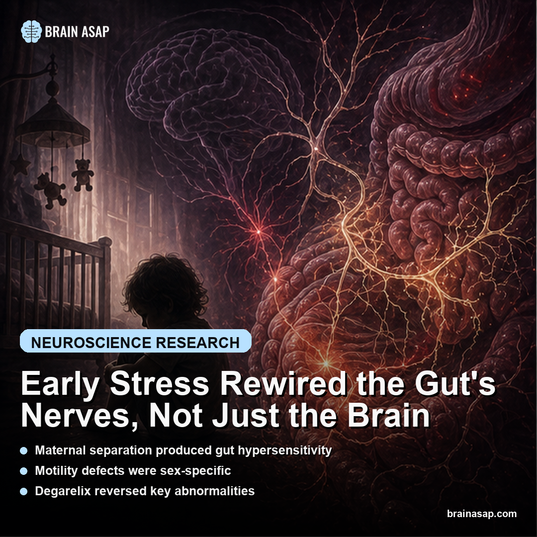
- Maternal separation produced gut hypersensitivity: The mouse model developed visceral pain sensitivity and lasting motility defects.
- Motility defects were sex-specific: The gut phenotype depended partly on sex hormones rather than being a single uniform stress effect.
- Degarelix reversed key abnormalities: Suppressing gonadal hormones reversed stress-induced visceral pain and motility defects in mice.
- Sympathetic input increased: Early stress enhanced sympathetic innervation of the enteric nervous system.
- Chemical sympathectomy restored motility: Reducing sympathetic drive normalized motility, pointing to a tractable neural pathway.
- Two pediatric cohorts aligned with the model: Maternal mental health problems were associated with pediatric disorders of gut-brain interaction.
Source: Gastroenterology (2026) | Najjar et al.
Early life stress is usually described as something that changes the brain. This paper makes the gut part of the same developmental pathway, showing how adversity can alter enteric neurons, sympathetic input, pain sensitivity, and motility long after the stressor ends.
Why the Gut-Brain Axis Needed a Nerve-Level Explanation
The phrase gut-brain axis is often used so broadly that it stops explaining anything. This paper gets more concrete. It asks which wires change after early adversity, which symptoms follow, and whether those pathways have human echoes.
In the maternal separation model, early stress did not just make animals broadly vulnerable. It produced visceral hypersensitivity, altered motility, and changes in the enteric nervous system, the nerve network embedded in the gut wall itself.
Sex Hormones Connected Early Stress to Adult Gut Motility
One important detail is that the motility defects were sex-specific. The reason is disorders of gut-brain interaction, including irritable bowel syndrome, often differ by sex in prevalence, symptom pattern, and clinical course.
The degarelix result sharpened the mechanism. By suppressing gonadal hormones, the researchers reversed stress-induced pain and motility changes. That does not make hormones the full explanation, but it places them inside the pathway from early adversity to adult gut function.
- Early stress input: maternal separation created a developmental stress exposure in mice.
- Gut output: the animals developed visceral pain sensitivity and altered motility.
- Hormone link: degarelix showed that gonadal hormone signaling contributed to the adult phenotype.
- Nerve link: sympathetic input to the enteric nervous system helped maintain the motility defect.
Why Sympathetic Overdrive Looks Like a Treatment Clue
The sympathetic nervous system is the body’s mobilization system. In the gut, too much sympathetic influence can disrupt the balanced choreography needed for normal motility. The paper found enhanced sympathetic innervation of the ENS after early stress.
Chemical sympathectomy restored motility in the mouse model, which is the kind of mechanistic rescue that changes interpretation. The gut problems were not only scars of development; they were maintained by a pathway that is plausibly manipulated.
Enteric Neurons Made the Stress-to-Gut Pathway More Concrete
Stress-related gut disorders are sometimes discussed as if the brain merely sends distress downward into the abdomen. The enteric nervous system changes that picture.
It is a dense neural network inside the gut wall, capable of coordinating motility, secretion, and local reflexes. If early stress reshapes that network, the gut is not only receiving a mood signal. It is developing differently.
The mouse data point to altered ENS composition, including changes in serotonergic innervation and subtype-specific neuronal proportions. That gives the paper a cellular substrate for long-term symptoms. Constipation, diarrhea, and visceral pain can then be understood as outputs of changed wiring rather than vague expressions of stress.
This is also why the human correlation is important. Pediatric disorders of gut-brain interaction are common and frustrating because structural tests often look normal. A developmental nerve-circuit model gives clinicians and researchers a more biologically specific way to think about symptoms that are real even when endoscopy is unrevealing.
How Sympathetic Drive Could Lock In a Childhood Signal
The sympathetic nervous system is supposed to mobilize the body during threat. In short bursts, that is adaptive. In a developing gut, persistent or exaggerated sympathetic influence may become a way for early stress to leave a lasting physiological fingerprint.
The study’s sympathetic finding is unusually actionable. Maternal separation enhanced sympathetic innervation of the ENS, and chemical sympathectomy restored motility.
That rescue suggests the adult phenotype was not simply a fixed developmental scar. It was at least partly maintained by an ongoing neural input.
The distinction is important for therapy. A fixed scar invites resignation; an active pathway invites intervention. The paper does not hand clinicians a treatment protocol, but it identifies sympathetic overactivity as a mechanistic target that is plausibly tested in future pharmacologic, neuromodulatory, or behavioral studies.
Why Human Cohorts Strengthen but Do Not Settle the Mechanism
The human side of the paper used two large pediatric cohorts to test whether early adversity signals, particularly maternal mental health problems, were associated with disorders of gut-brain interaction. That is a sensible translational bridge because the mouse model cannot capture the complexity of childhood family stress, medical access, diet, genetics, and reporting bias.
The cohort result does not prove that a child’s gut symptoms are caused by the same enteric and sympathetic mechanisms observed in mice. Human adversity is messy, and gut disorders are heterogeneous. But seeing the association in both directions, mechanistic in animals and epidemiologic in children, makes the result harder to dismiss.
The most careful interpretation is that early stress can shape gut-brain biology through multiple routes. This paper names several of them: enteric neuronal composition, sex-hormone-sensitive motility, sympathetic innervation, and visceral sensory pathways. That is a much better starting point than saying stress affects the gut and stopping there.
What Would Make the Human Translation Stronger
The next human step is not just showing that adversity predicts pediatric gut symptoms. It is measuring the intermediate biology. Autonomic tone, stress hormones, inflammatory markers, sex-hormone status, motility testing, and noninvasive proxies of enteric function can help connect the mouse pathway to children with disorders of gut-brain interaction.
Longitudinal timing will matter too. If early adversity changes the gut during a sensitive developmental window, then symptoms can emerge only when later hormones, infections, diet, or stress load push the system. A cohort that follows children over time could reveal whether sympathetic markers appear before motility and pain problems or only after symptoms begin.
The therapeutic possibility is not to erase childhood stress from biology. It is to identify the active pathways that keep amplifying symptoms. This paper makes sympathetic tone, enteric neuron composition, and hormone-sensitive motility worth watching closely.
Functional Gut Symptoms Need Positive Biology
Functional gut disorders are often described by what they are not: not cancer, not inflammatory bowel disease, not an ulcer. This paper gives a better positive language. It points to altered enteric wiring, sympathetic overdrive, sex-hormone-sensitive motility, and visceral sensory amplification.
The biology can reduce stigma. If early stress can change the systems that govern pain and motility, then symptoms are not imaginary just because routine structural tests are normal. The gut can be mechanically intact and neurologically dysregulated at the same time.
Pediatric Gut Pain Can Carry a Developmental Signal
The human cohorts do not prove the mouse pathway operates identically in children. They show that maternal mental health and early adversity track with pediatric disorders of gut-brain interaction, giving the animal mechanism a clinically recognizable frame.
The precision is the advance. Instead of treating stress-related gut disorders as vague psychosomatic spillover, the paper points to enteric composition, sex hormones, and sympathetic innervation as biological targets worth testing.
Paper: Enteric and Sympathetic Nervous System Pathways Mediate Early Life Stress Effects on Gut Motility and Pain: Mechanistic Findings With Human Correlation. Gastroenterology. 2026. DOI: 10.1053/j.gastro.2026.02.030
Authors: Najjar et al.
Study Design: Maternal-separation mouse model with mechanistic interventions plus analysis of two pediatric population cohorts.
Sample Size: Mouse model experiments and two large human pediatric cohorts.
Key Statistic: Chemical sympathectomy restored motility after early-life-stress-induced sympathetic innervation of the ENS.