
Early Locus Coeruleus Axon Loss May Explain Smell Loss in Alzheimer’s
TL;DR: Alzheimer’s-model mice lost noradrenergic locus coeruleus axons in the olfactory bulb before major plaque buildup, took 60% longer to find buried food, and improved when microglial phagocytosis was reduced.
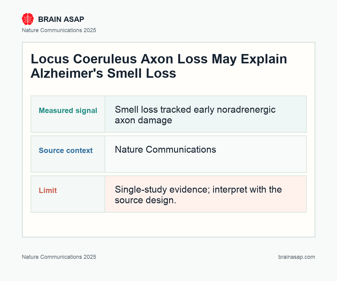
Key Findings
- 3-month axon loss before heavy plaque load: AppNL-G-F mice showed a selective drop in noradrenergic fibers in the olfactory bulb at an early age, while cholinergic and serotonergic neurites were preserved.
- 60% slower buried-food performance: At 3 months, the Alzheimer’s-model mice needed about 60% more time to find buried food, linking axon loss to a measurable hyposmia-like phenotype.
- 33% higher microglial phagocytosis: Primary olfactory-bulb microglia from AppNL-G-F mice showed a 33% increase in phagocytic activity, and microscopy found more locus coeruleus axon material inside microglial lysosomes.
- TSPO knockout rescued smell behavior: Cutting phagocytosis via TSPO deletion preserved locus coeruleus axons and restored buried-food performance toward wild-type levels.
- LC-restricted mutant APP was enough: Driving human AppNL-G-F only in locus coeruleus neurons still caused roughly 15% olfactory-bulb axon degeneration and a matching smell deficit.
- Human translation signal appeared early: TSPO-PET showed elevated olfactory-bulb signal in 16 prodromal AD patients versus 14 controls, and post-mortem early-AD olfactory bulbs showed reduced locus coeruleus axon density.
Source: Nature Communications (2025) | Meyer et al.
Smell loss often shows up before memory failure in Alzheimer’s disease, but that early sensory change has been oddly hard to pin to a mechanism. This paper lands on a surprisingly specific answer: the problem may begin when locus coeruleus axons into the olfactory bulb are stripped away early by microglia, long before classic plaques dominate the picture.
Why Early Smell Loss in Alzheimer’s Has Always Felt Mechanistically Awkward
Olfactory decline is one of the most repeated “early sign” claims in Alzheimer’s disease, yet the field has struggled to explain it cleanly. Plaques and tangles show up in many regions.
Smell is processed across several stations. And most people with emerging smell problems do not get an immediate mechanistic account, just a vague warning that something neurodegenerative may be brewing.
This paper makes that mechanism much more specific. The authors focused on the locus coeruleus, the noradrenergic brainstem hub that modulates arousal, attention, and sensory processing, including input into the olfactory bulb.
That region is already known to be hit early in Alzheimer’s biology. What was missing was proof that its axons into the olfactory bulb degenerate early enough, and locally enough, to help explain smell loss before overt dementia.
Their answer was yes, and the mechanism was not passive degeneration alone. Microglia appeared to be actively clearing those axons.
What Happened in the Olfactory Bulb Before Plaques Took Over
In AppNL-G-F mice, the team found early loss of noradrenergic fibers in the olfactory bulb, with the most prominent hit in the internal plexiform layer. This stood out because cholinergic and serotonergic neurites were not reduced at the same age. The damage looked selective, not like a generic collapse of every modulatory input.
Behavior lined up with anatomy. At 3 months, the transgenic mice needed about 60% more time to find buried food, a classic olfactory task.
In a second vanilla-odor test, they also showed reduced investigatory behavior. The combination is strong: structural loss in the sensory circuit, impaired performance on two smell-relevant tasks, and reduced odor-evoked noradrenaline release in the olfactory bulb.
The smell circuit was not just missing fibers on a slide. It was functionally under-supplied by a brainstem system that normally helps tune sensory processing, attention, and arousal.
How Microglia Turned a Vulnerable Circuit Into a Removable Target
Here, the paper gets much more interesting than a standard pathology study. Bulk RNA sequencing and functional assays suggested that olfactory-bulb microglia were not just present in larger numbers.
They were more phagocytic. In culture, microglia from AppNL-G-F mice showed a 33% higher ability to engulf labeled synaptosomes.
Microscopy pushed the mechanism further. The team found more noradrenergic axon material inside microglial lysosomes, plus evidence that those axons were exposing phosphatidylserine, the classic “eat me” signal. MFG-E8 appeared to act as the adaptor linking that externalized phosphatidylserine to microglial engulfment machinery.
That turns the interpretation from one of simple axon fragility into one of active removal. Hyperactive or stressed locus coeruleus axons in the olfactory bulb may be getting tagged for clearance, then physically eaten by microglia before large-scale amyloid pathology dominates the region.
- Axon signal: noradrenergic fibers were selectively reduced in the olfactory bulb.
- Microglial signal: AppNL-G-F microglia showed higher phagocytic activity and contained more axon material.
- Rescue signal: reducing phagocytosis through TSPO deletion preserved axons and improved buried-food performance.
The Rescue Experiments Strengthened the Locus Coeruleus Claim
Correlation alone would not have been enough. The strongest evidence in the paper comes from the rescue experiments. When the authors crossed the Alzheimer’s-model mice with TSPO-knockout animals, they reduced microglial phagocytosis, preserved more locus coeruleus axons, and rescued buried-food performance at 3 months.
They also ran the inverse logic test. Restricting human AppNL-G-F expression specifically to locus coeruleus neurons still caused about 15% axon degeneration in the olfactory bulb and produced a smell deficit.
That narrows the causal chain. You do not need whole-brain transgenic pathology to get the phenotype. Hitting the locus coeruleus projection alone was enough to reproduce a smaller version of the problem.
The rescue and restriction experiments make the mechanism harder to explain as a generic plaque side effect. They point to an early circuit-specific process: vulnerable noradrenergic axons are marked, cleared, and then smell behavior suffers.
How the Human Data Push This Beyond a Mouse Curiosity
The human arm was not huge, but it mattered. The group used TSPO-PET in 16 people with prodromal Alzheimer’s biology, 16 with AD, and 14 unaffected controls. The olfactory bulb showed elevated TSPO signal already in the prodromal group, suggesting increased microglial burden or activity before later-stage diagnosis added much more signal.
They also examined post-mortem olfactory bulbs from early-AD donors and found reduced locus coeruleus axon density, matching the broad direction of the mouse work. Human olfactory testing in the prodromal group only showed a trend rather than a clean slam-dunk deficit, and the authors acknowledge the small cohort size as a limitation. Still, the translational bridge is unusually strong for this kind of circuit paper.
The human data also keep the mouse result from becoming too neat. TSPO-PET is a proxy for glial biology, not a direct movie of microglia eating axons, and smell loss in people has many causes.
The key clinical question is whether this circuit can be measured early enough to change care. A smell test alone is too nonspecific; allergies, infection, aging, Parkinson’s disease, and medication effects can all muddy the signal. But pairing olfactory testing with TSPO-PET, locus-coeruleus-sensitive imaging, or fluid markers of microglial activity could turn a vague early warning sign into a more biologically anchored risk marker.
This study suggests that one of Alzheimer’s earliest outward signs may be less about the nose itself and more about an early loss of noradrenergic support to the olfactory bulb. If that holds up, smell testing plus targeted imaging of this circuit could become a more biologically specific early-detection strategy.
Paper: Early Locus Coeruleus noradrenergic axon loss drives olfactory dysfunction in Alzheimer’s disease. Nature Communications. 2025;16:7338.. DOI: 10.1038/s41467-025-62500-8
Authors: Meyer et al.
Study Design: Cohort study
Sample Size: Human translation signal appeared early: TSPO-PET showed elevated olfactory-bulb signal in 16 prodromal AD patients versus 14 controls, and post-mortem early-AD olfactory bulbs showed reduced locus coeruleus axon density.
Key Statistic: 3-month axon loss before heavy plaque load: AppNL-G-F mice showed a selective drop in noradrenergic fibers in the olfactory bulb at an early age, while cholinergic and serotonergic neurites were preserved.