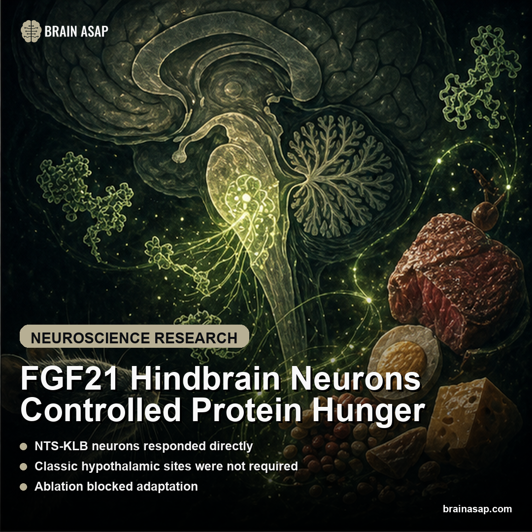
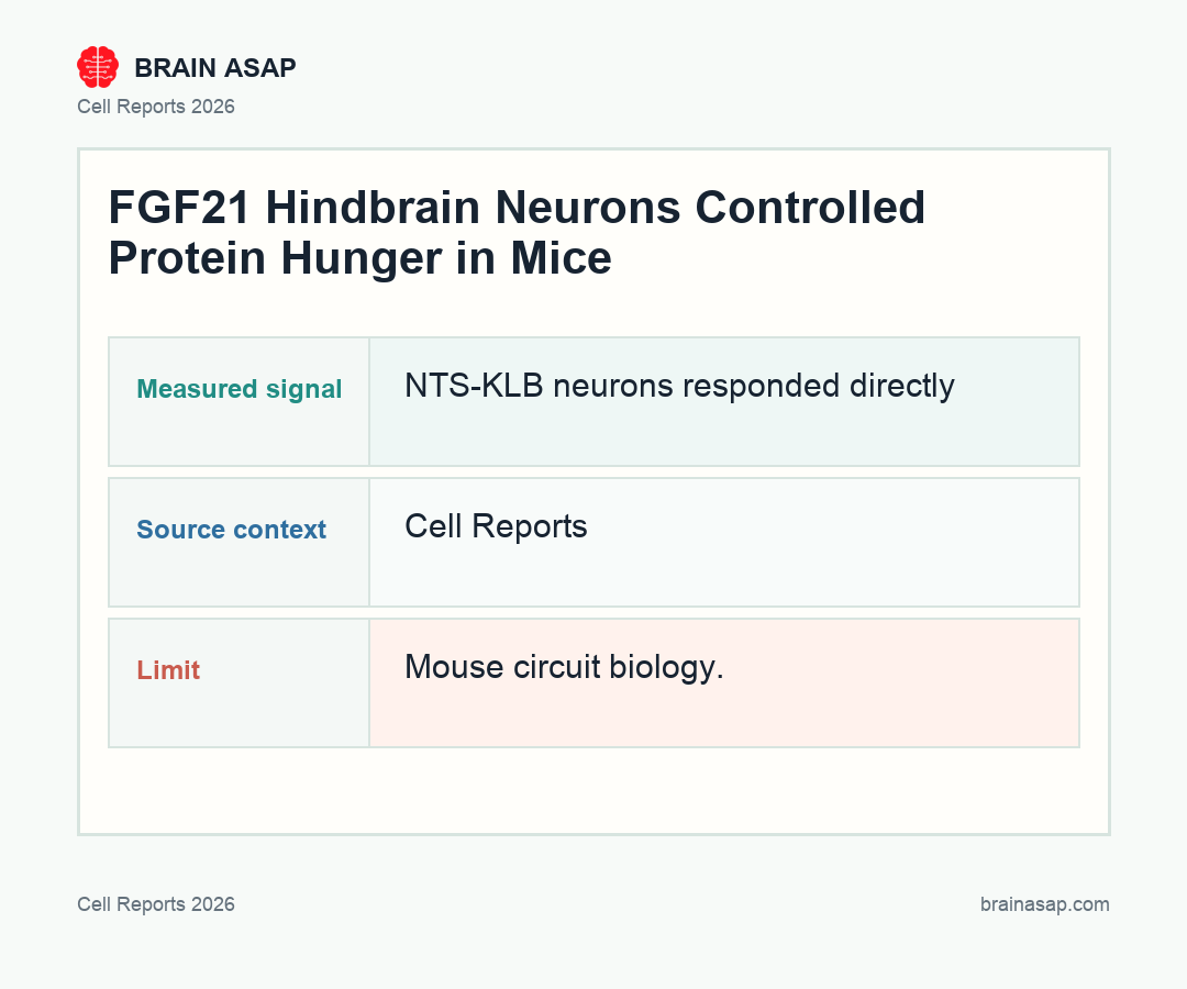
FGF21 Hindbrain Neurons Controlled Protein Hunger
TL;DR: During protein restriction, liver-derived FGF21 acted through glutamatergic beta-klotho neurons in the nucleus of the solitary tract to change food intake, food choice, and energy expenditure in mice.
Key Findings
- NTS-KLB neurons responded directly: Klb-expressing glutamatergic neurons in the nucleus of the solitary tract were activated by FGF21.
- Classic hypothalamic sites were not required: Systematic tests found the SCN, PVN, and VMH were not required for the FGF21-mediated responses examined.
- Ablation blocked adaptation: Selective ablation of NTS-KLB neurons prevented changes in food intake, food choice, and energy expenditure during protein restriction.
- Activation was sufficient: Chemogenetic activation of those neurons was sufficient to drive the adaptive responses.
- Circuit links diet to brainstem control: The findings identify a neural pathway connecting dietary protein sensing to metabolic adaptation.
Source: Cell Reports (2026) | Spann et al.
FGF21 is often described as a metabolic hormone, but hormones need circuits. This Cell Reports study identifies a specific hindbrain population that translates a low-protein diet into behavior and energy use.
The Liver Signal Needed a Hindbrain Receiver
When protein intake drops, the liver releases FGF21. That hormone tells the body something important: the diet is missing a key macronutrient, and behavior should change.
The question was where that message becomes a neural command. The answer in this mouse study was a discrete population of beta-klotho-expressing glutamatergic neurons in the nucleus of the solitary tract, or NTS.
Beta-klotho is part of the receptor machinery FGF21 needs to act on cells. Finding Klb-expressing neurons in the NTS gave the authors a concrete target instead of a vague brainwide hormone effect.
- Signal source: Protein restriction increased the liver-derived FGF21 message.
- Brain receiver: NTS neurons expressing beta-klotho responded to that hormonal signal.
- Behavioral output: The circuit shaped intake, food choice, and energy expenditure during protein restriction.
The Hypothalamus Was Not the Required Control Center
Metabolic control is often routed through hypothalamic regions such as the suprachiasmatic nucleus, paraventricular nucleus, and ventromedial hypothalamus. The authors tested those candidates directly.
In this context, they were not required for FGF21-mediated responses to protein restriction. That result pushes the spotlight to the hindbrain, a region sometimes treated as more basic but deeply involved in appetite, visceral sensing, and energy balance.
The result revises the usual map of appetite control. The hypothalamus is central, but it is not the only place where metabolic information becomes behavior.
The NTS is well positioned for this job because it integrates visceral information, circulating signals, and feeding-related control. A liver hormone reaching a hindbrain receiver is therefore a plausible route for changing the animal’s response to a low-protein diet.
The finding also narrows the therapeutic question. If FGF21 drugs change eating behavior, the relevant circuitry may include brainstem nodes that are often missed when research looks only at hypothalamic appetite centers.
Ablation and Activation Made the Circuit Convincing
The logic was two-sided. Remove NTS-KLB neurons, and mice failed to mount normal metabolic adaptations to protein restriction. Activate them, and the responses appeared.
That necessary-and-sufficient structure gives the paper its strength. The neurons were not just active during the behavior. They were functionally positioned to coordinate it.
Many circuit studies stop at correlation: a population lights up during a behavior, so it becomes a candidate node. This paper went further by perturbing the cells and watching whether the adaptation survived.
Ablation tested whether the cells were required. Chemogenetic activation tested whether stimulating them could recreate the adaptive pattern. Together, those experiments make a stronger case that NTS-KLB neurons sit inside the control pathway.
The work also links several outputs that are often studied separately. Intake, macronutrient choice, and energy expenditure are coordinated responses, not isolated dials.
Protein Appetite Is a Brain-Body Negotiation
The study makes protein hunger feel less like willpower and more like physiology. A liver hormone reports dietary state, a hindbrain circuit receives the signal, and the animal changes intake, food choice, and energy expenditure.
The circuit matters for obesity and metabolic disease research because FGF21-based therapies are already being explored clinically. Understanding the route can help define endpoints beyond liver fat, including dietary behavior and metabolic rate.
This is also a reminder that hunger is not one sensation. Protein need, energy need, gut state, reward value, and learned food preference can pull behavior in different directions.
FGF21 appears to be one way the body communicates a specific nutritional problem. The signal is not simply “eat more” or “eat less”; it helps redirect physiology during a low-protein state.
The distinction affects treatment development. A drug that changes weight without clarifying food choice, protein appetite, or energy expenditure may miss the mechanism behind durable benefit or risk.
The NTS finding also makes the brainstem feel less like a relay station and more like an active decision point. The nucleus of the solitary tract receives visceral information from the gut and body, so it is well placed to compare what the animal ate, what the liver is reporting, and what behavior should change next.
Protein restriction therefore produced a coordinated package rather than one isolated readout. Intake, preference, and energy expenditure all shifted because the animal was solving a nutritional imbalance, not just responding to a single appetite cue.
For human metabolism, the distinction is clinically important. Many weight-loss and diabetes interventions change appetite, nausea, food reward, and energy balance at once. A circuit-level map can help researchers tell which effects are part of adaptive nutrient sensing and which are side effects of pushing the system too hard.
Protein-Hunger Therapy Still Needs Human Testing
This is mouse circuit biology. It does not say that targeting NTS-KLB neurons in humans is ready or even straightforward.
But it does sharpen the map. If FGF21 treatments work partly through brain circuits, then the next generation of metabolic therapy needs to measure not just blood chemistry but how hunger, food choice, and energy expenditure are being neurally coordinated.
Human studies would need careful endpoints. Protein preference, total intake, cravings, body composition, metabolic rate, nausea, and reward-related eating could all change in clinically important ways.
They would also need to separate protein hunger from general appetite suppression. A therapy that reduces calories while worsening protein adequacy would not be the same as a therapy that helps the body correct a macronutrient imbalance.
That is why this circuit study is useful even before translation. It gives researchers a cleaner vocabulary for asking what kind of hunger is changing.
It also suggests that diet composition should be measured with more care in metabolic trials, not treated as background noise.
Protein appetite is a specific biological problem, and this paper finally gives it a specific circuit address.
That address should make future FGF21 studies much harder to keep vague for clinicians and trialists.
The translational opportunity is not to manipulate a tiny brainstem population directly. It is to understand how liver signals, diet composition, and brain circuits cooperate when the body detects nutritional imbalance.
Metabolism is not peripheral to the brain in this model. The brain helps decide what the body does with a dietary problem, and hormones like FGF21 are part of that control system.
Paper: FGF21 signals through hindbrain neurons to alter food intake and energy expenditure during dietary protein restriction. Cell Reports. 2026. DOI: 10.1016/j.celrep.2026.117218
Authors: Spann et al.
Study Design: Preclinical mouse circuit study using Klb-Flp intersectional genetics, neuronal ablation, chemogenetic activation, and metabolic/behavioral assays.
Sample Size: Mice across protein-restriction, neuronal ablation, and chemogenetic activation experiments; exact group counts should be checked in the full paper before publication.
Key Statistic: NTS beta-klotho neurons were directly activated by FGF21, required for protein-restriction adaptations, and sufficient to drive changes in food intake, food choice, and energy expenditure.