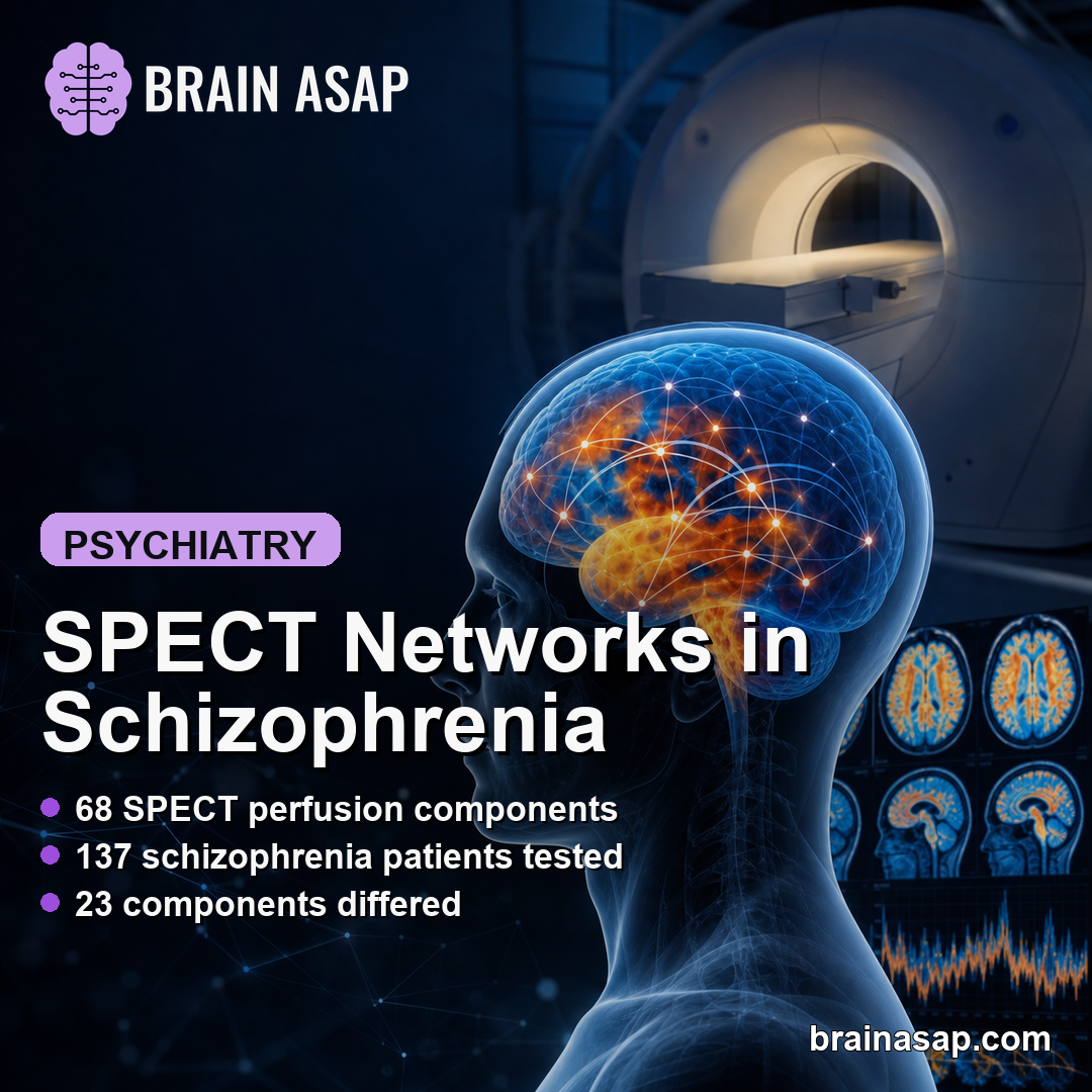
TL;DR: A 2026 medRxiv preprint introduced a 68-component NeuroMark template for single photon emission computed tomography (SPECT), a brain blood-flow imaging method, and found schizophrenia-related differences in cerebellar, subcortical, higher-cognition, visual, and triple-network perfusion patterns.
Key Findings
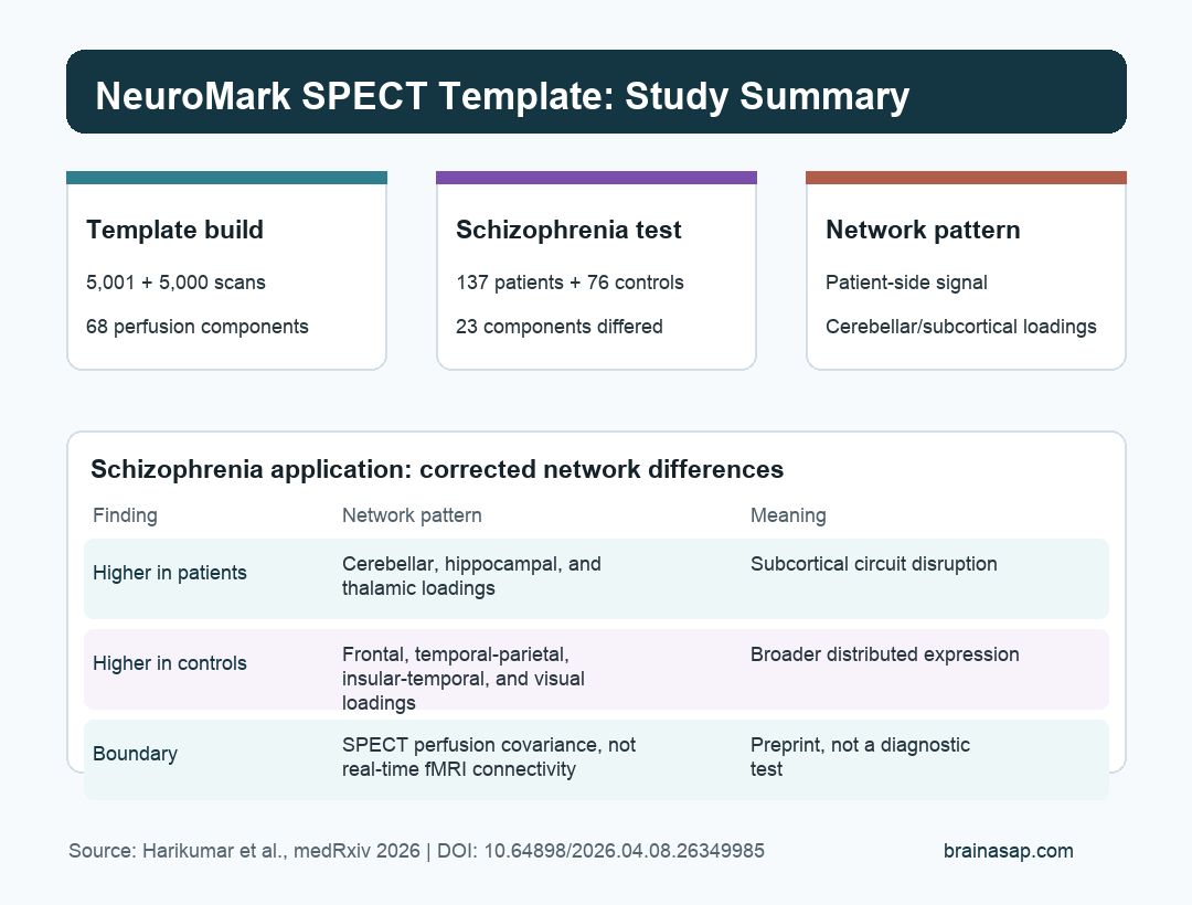
- Template build: Researchers used blind independent component analysis on two large SPECT datasets of 5,001 and 5,000 scans to identify repeatable perfusion components.
- Final template: After quality control and cross-run matching, the new NeuroMark SPECT template retained 68 brain perfusion components.
- Schizophrenia test set: The template was then applied to 137 schizophrenia patients and 76 healthy controls.
- Loading differences: 23 of 68 components differed after false-discovery-rate correction, with patient-side loadings strongest in cerebellar and subcortical domains.
- Clinical boundary: The work is a preprint and should be read as a research framework, not a clinical SPECT test for diagnosing schizophrenia.
Source: Harikumar et al. medRxiv. 2026.
SPECT brain scans estimate regional cerebral blood flow. In psychiatric imaging research, that gives researchers one way to study whether brain perfusion patterns organize into repeatable networks.
The Harikumar preprint focused on a missing tool. Functional MRI has NeuroMark templates that help researchers compare brain network components across studies, but SPECT has not had a comparable template for automated network analysis.
Researchers built one, then tested whether it could detect schizophrenia-related differences in a separate clinical imaging cohort.
Researchers Built a 68-Component SPECT Perfusion Template
The team started with two large SPECT datasets. Blind independent component analysis, or blind ICA, was run separately on 5,001 scans in one round and 5,000 scans in a second round.
Independent component analysis separates a complex image dataset into spatial patterns that covary across people. Here, those patterns represented SPECT perfusion components, not real-time neural communication.
After automated labeling, visual inspection, and cross-run matching, researchers kept 68 replicated components. Components that looked noisy, white-matter-like, artifactual, or weakly matched between runs were excluded.
- Input modality: SPECT scans were used to estimate resting cerebral blood flow.
- Template goal: The template was meant to make SPECT network estimation more comparable across people and studies.
- Component domains: The retained components were organized into cerebellar, higher-cognition, subcortical, sensorimotor, triple-network, and visual domains.
- Research output: The template can be used with spatially constrained ICA to estimate participant-level perfusion network expression.
A template changes the analysis from a one-off decomposition into a reusable reference. Future studies can ask whether the same component space carries different clinical information in different cohorts.
Schizophrenia Patients and Controls Were Tested Separately
To show how the template works, researchers applied it to an independent schizophrenia dataset from Amen Clinics. The test cohort included 137 schizophrenia patients and 76 healthy controls.
The analysis used spatially constrained ICA, abbreviated sc-ICA. The new template acted as a guide while the algorithm estimated how strongly each component appeared in each participant.
Researchers then compared two related measurements:
- Loading parameters: How strongly each of the 68 components was expressed in patients versus controls.
- Co-modulation matrices: How component expression patterns covaried with each other across the template.
Those measurements do not say that one region is simply “on” or “off.” They describe the strength and covariance of SPECT perfusion patterns across network-like components.
Twenty-Three SPECT Components Differed After Correction
The component-loading comparison produced the main numeric result. After false-discovery-rate correction, 23 of 68 components differed between schizophrenia patients and healthy controls.
Five components showed higher loading expression in patients. Eighteen components showed higher loading expression in controls.
The patient-side pattern was concentrated in cerebellar, subcortical extended hippocampal, and subcortical extended thalamic domains. The control-side pattern was broader, with higher loadings across higher-cognition frontal, temporoparietal, insular-temporal, default-mode, and visual-occipital domains.
- Patient-side loadings: Stronger expression appeared in cerebellar and subcortical components, including extended hippocampal and thalamic domains.
- Control-side loadings: Stronger expression appeared across higher-cognition and visual components.
- Temporal lobe note: The preprint highlighted increased temporal-lobe loading in one component as potentially relevant to schizophrenia symptoms such as auditory hallucinations.
- Statistical guardrail: The 23-component result used false-discovery-rate correction, which reduces the chance of treating random component differences as meaningful.
The pattern fits a familiar schizophrenia imaging theme: disruption is not limited to one isolated region. It can involve distributed cerebellar, subcortical, cognitive-control, default-mode, and sensory networks.
Co-Modulation Suggested More Concentrated Patient-Side Network Coupling
The co-modulation analysis asked a different question. Instead of testing component strength one at a time, it examined how component patterns covaried across the template.
Controls showed a more distributed co-modulation pattern across cognitive, executive, visual, and default-mode systems. Patients showed a more concentrated pattern, especially involving subcortical, cerebellar, visual, and higher-cognition domains.
The study authors interpreted these differences as consistent with altered cerebello-thalamocortical and visual circuitry in schizophrenia. That interpretation fits prior work linking schizophrenia to changes in default-mode, triple-network, sensory, and cognitive-control systems.
Co-modulation should not be confused with fMRI functional connectivity. SPECT-derived co-modulation reflects spatial covariance in perfusion components, not second-by-second synchrony between brain areas.
The Template Is Useful, But the Clinical Claim Should Stay Narrow
The study’s most durable contribution may be methodological. A shared SPECT template could make future perfusion-network studies easier to compare, especially when researchers want participant-specific component estimates.
The schizophrenia finding is still preliminary. The preprint was not peer reviewed at the time of posting, the control group was smaller than the patient group, and the data came from a clinical imaging setting rather than a population-based cohort.
- Preprint status: The findings should not guide clinical care until they survive peer review and independent replication.
- Sample imbalance: The test cohort had 137 patients and 76 controls, which is workable for demonstration but not definitive.
- Modality boundary: SPECT perfusion covariance is not the same measurement as fMRI connectivity or structural MRI.
- No diagnostic endpoint: The analysis did not validate a clinical SPECT screen for schizophrenia.
Harikumar et al. built a replicable NeuroMark SPECT template, then showed that it can detect interpretable schizophrenia-related perfusion-network differences.
The work is an imaging-methods step. It does not turn SPECT into a stand-alone diagnostic tool.
Citation: DOI: 10.64898/2026.04.08.26349985. Harikumar et al. A Replicable NeuroMark Template for Whole-Brain SPECT Reveals Data-Driven Perfusion Networks and Their Alterations in Schizophrenia. medRxiv. 2026.
Study Design: Neuroimaging methods preprint using blind ICA template construction and spatially constrained ICA application in an independent schizophrenia cohort.
Sample Size: Template construction used two SPECT datasets of 5,001 and 5,000 scans; the schizophrenia application used 137 patients and 76 controls.
Key Statistic: 23 of 68 NeuroMark SPECT components differed between schizophrenia patients and controls after false-discovery-rate correction.
Caveat: The source is a preprint and the results are research findings, not a validated clinical diagnostic test.