TL;DR: A 2026 study in Scientific Reports identified a novel OPHN1 K306N variant in a boy with cyclic strabismus, linking a 24-hour eye-alignment rhythm to altered phosphoinositide binding without the intellectual disability usually associated with OPHN1 syndrome.
Key Findings
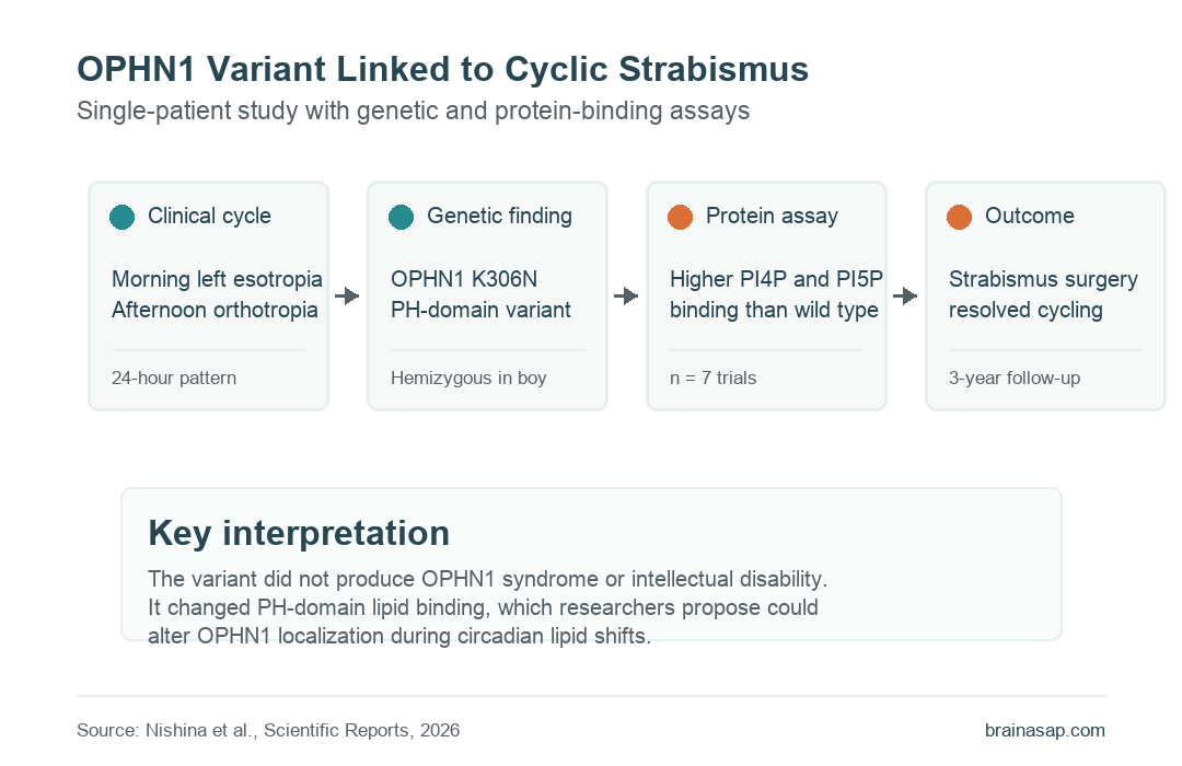
- 8-year-old patient: A Japanese boy had cyclic esotropia, with left eye crossing in the morning and normal alignment later the same day.
- 24-hour cycle: Calendar tracking and repeated examinations showed alternating esotropia and orthotropia across half-day periods.
- OPHN1 K306N variant: Trio whole-exome sequencing found a hemizygous c.918G>T, p.Lys306Asn variant in the OPHN1 PH domain.
- No OPHN1 syndrome: The child had normal psychomotor development, no intellectual disability, and no other neurological or systemic abnormalities.
- Higher lipid binding: Protein assays found that OPHN1 K306N increased binding to PI4P and PI5P compared with wild-type OPHN1.
Source: Scientific Reports (2026) | Nishina et al.
Cyclic strabismus is a rare eye-alignment disorder in which periods with and without strabismus alternate in a rhythmic pattern. The unusual part of this case was the link between that cycle and a gene better known for neurodevelopmental disease.
OPHN1 encodes oligophrenin-1, a Rho GTPase-activating protein involved in cell signaling and morphology. Pathogenic OPHN1 mutations can cause intellectual disability, seizures, hypotonia, abnormal behavior, and strabismus, but this patient did not have that broader syndrome.
A Boy Had Morning Esotropia and Afternoon Orthotropia
The patient was born at 40 weeks and had normal psychomotor development. At age 5, he suddenly developed left-eye esotropia and double vision, but the eye crossing resolved spontaneously and then recurred occasionally.
By age 8, his mother recorded days and half-days with and without eye crossing on a calendar. The pattern led clinicians to diagnose cyclic esotropia with a 24-hour cycle.
- Morning examination: The left eye showed constant esotropia of 40 prism diopters at near and far distances.
- Afternoon examination: The same day, the patient showed orthotropia, meaning normal eye alignment, with fine stereoacuity of 40 seconds.
- Brain imaging: Head MRI found no abnormality that explained the eye-alignment cycle.
Strabismus surgery later corrected the alignment. After left medial rectus recession and left lateral rectus resection, the patient maintained orthophoria and had no recurrence across 3 years of follow-up.
Whole-Exome Sequencing Found OPHN1 K306N
Researchers used trio-based whole-exome sequencing (WES), a genetic test that reads the protein-coding regions of the patient and both parents. The analysis found a hemizygous OPHN1 missense variant in the patient and a heterozygous variant in his mother.
The variant was c.918G>T, p.Lys306Asn, also written as K306N. It sits in the PH domain, a protein region that can bind phospholipids and influence where a protein localizes inside the cell.
- Database absence: The variant was absent from gnomAD and the Tohoku Medical Megabank population reference.
- Conserved residue: Lysine at position 306 was conserved across human, marmoset, mouse, and zebrafish OPHN1.
- Prediction scores: In silico tools suggested possible damage, including SIFT 0.0418, PolyPhen-2 0.991, and CADD 26.2.
The authors still classified the variant as a variant of uncertain significance under ACMG guidelines. One case cannot prove causation by itself.
OPHN1 K306N Increased PI4P and PI5P Binding
The team used AlphaFold 3 to model how the OPHN1 PH domain might bind phosphatidylinositol 4,5-bisphosphate, a membrane lipid. The model placed K306 near the phospholipid-binding pocket.
Researchers then purified HA-tagged wild-type and K306N OPHN1 proteins from HEK293T cells and tested binding to phosphoinositide-containing liposomes. The experiment focused on PI3P, PI4P, and PI5P.
- PI3P: Wild-type and K306N OPHN1 did not differ meaningfully in binding.
- PI4P: The K306N variant showed enhanced binding compared with wild-type OPHN1.
- PI5P: The K306N variant also showed enhanced binding compared with wild-type OPHN1.
- Replication: The blot result was described as representative of 7 trials.
The authors interpreted K306N as a gain-of-function change in the PH domain. In this context, gain of function means the altered protein binds certain membrane lipids more strongly, not that it improves eye movement.
A Circadian Lipid-Localization Hypothesis Could Explain the Cycle
The proposed mechanism is still hypothetical. The authors suggested that circadian rhythm-dependent changes in local PI4P or PI5P production could pull OPHN1 K306N toward those lipids and away from its usual cellular location.
If OPHN1 shifts location cyclically, local Rho signaling could change in a rhythmic way. That could produce temporary neurophysiological dysfunction in eye-alignment pathways, creating alternating periods of esotropia and normal alignment.
- Why OPHN1 fits: OPHN1 is linked to Rho signaling, synaptic function, hippocampal circadian-clock interactions, and neurodevelopmental phenotypes.
- Why this case differs: The K306N variant affected the PH domain, while many OPHN1 syndrome mutations affect other domains and produce broader neurodevelopmental symptoms.
- What remains unknown: The study did not show the variant changing OPHN1 localization in patient neurons or directly causing the circadian eye cycle.
The important result is therefore narrow: a rare OPHN1 PH-domain variant appeared in a child with cyclic strabismus and changed protein-lipid binding in vitro.
OPHN1 Findings Need More Cyclic Strabismus Cases
This study gives cyclic strabismus a plausible molecular lead, but it does not establish OPHN1 K306N as a confirmed cause. The evidence comes from one patient, inherited carrier status in the mother, prediction tools, structural modeling, and protein-binding assays.
Future work would need additional patients, functional studies in relevant eye-movement or neuronal models, and direct tests of whether circadian lipid shifts change OPHN1 localization. For now, the case suggests that some cyclic strabismus may involve a gain-of-function OPHN1 mechanism rather than the classic OPHN1 syndrome pathway.
Citation: DOI: 10.1038/s41598-026-48129-7. Nishina et al. A novel OPHN1 variant associated with cyclic strabismus but in the absence of OPHN1 syndrome. Scientific Reports. 2026;16:12200.
Study Design: Single-patient clinical genetics and protein-function study using trio whole-exome sequencing, structural prediction, and phosphoinositide-binding assays.
Sample Size: One 8-year-old boy with cyclic esotropia, plus parental genetic testing and OPHN1 protein assays in cultured cells.
Key Statistic: OPHN1 K306N increased binding to PI4P and PI5P compared with wild-type OPHN1, while the clinical cycle resolved after surgery with no recurrence over 3 years.
Caveat: The variant remains a single-case candidate mechanism; direct causation and circadian localization changes were not proven in patient neurons.