How Parasites Hack Your Gut-Brain Connection
TL;DR: Parasitic infections trigger a sophisticated cellular conversation in the gut that signals the brain to stop eating—a protective response that reveals how infection hijacks the gut-brain axis through epithelial cell crosstalk, acetylcholine release, and serotonin signaling.
When you’re fighting an infection, appetite disappears. It feels intuitive—your body’s way of conserving energy for immune defense. But the mechanism behind this response is far stranger than simple malaise. A new study reveals that parasites don’t just cause inflammation; they unlock a hidden communication circuit between specialized gut cells and the brain that systematically suppresses hunger.
Key Findings
- Parasites activate tuft-EC cell crosstalk: Helminth infection triggers direct communication between tuft cells and neighboring epithelial cells (EC cells), a previously unknown cellular interaction that initiates the entire gut-brain response.
- Acetylcholine release occurs in two phases: Tuft cells first release ACh through an unconventional non-synaptic mechanism in response to parasite metabolites, followed by sustained release during type 2 inflammation.
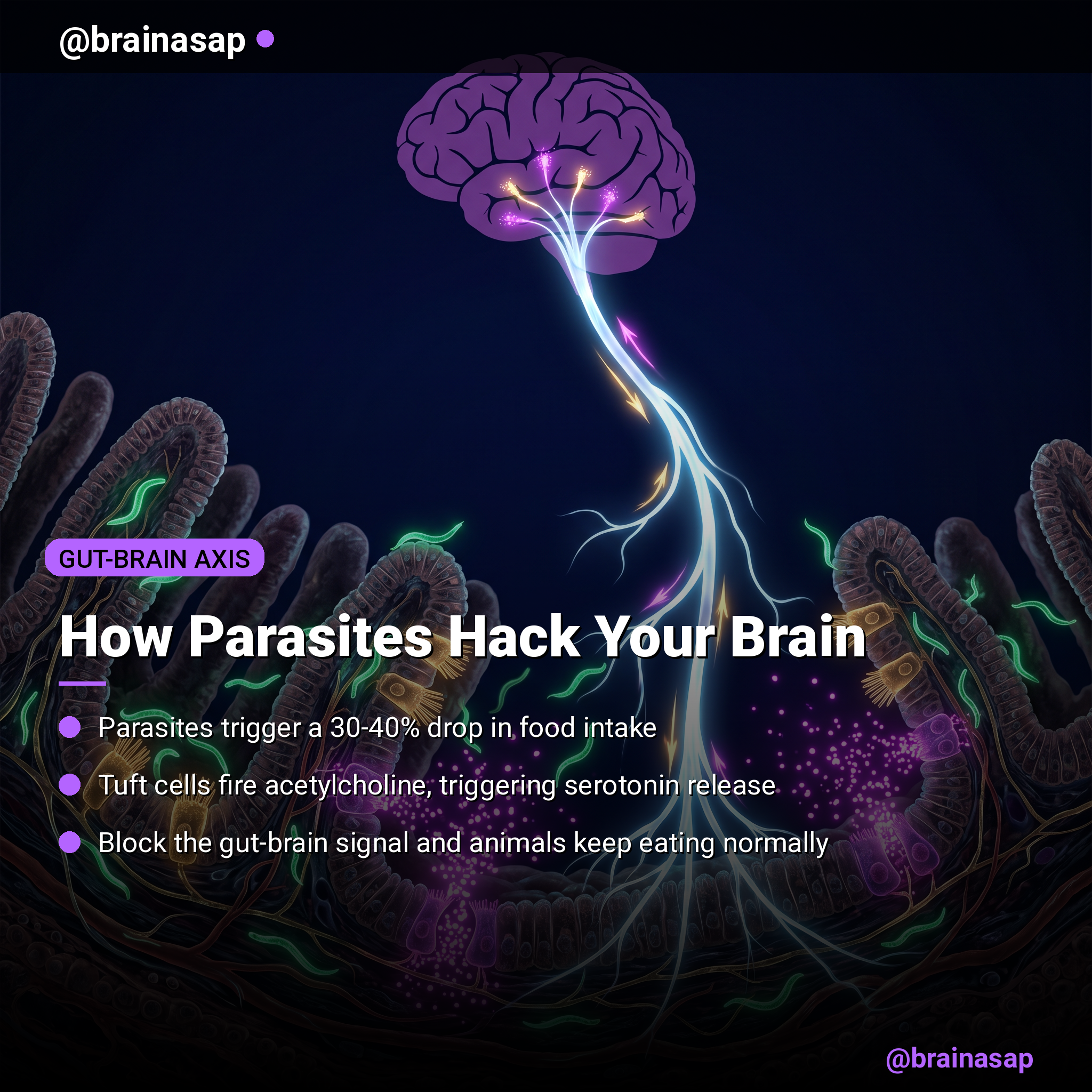
- Serotonin amplifies the signal: EC cells respond to ACh by releasing serotonin, which activates vagal nerve fibers that directly communicate with appetite-suppressing circuits in the brainstem.
- Food intake drops significantly: Infected mice showed 30% to 40% reduction in food intake during type 2 immune responses, an effect completely blocked when tuft-EC communication was prevented.
- Vagal signaling is essential: Severing vagal nerve communication or removing serotonin signaling eliminated the appetite-suppressing effect, confirming that gut-brain signaling is absolutely required for the response.
- The pathway activates during parasitic disease progression: Early parasite metabolites trigger acute acetylcholine release; sustained inflammation later maintains the signal, creating a coordinated multi-stage mechanism.
Source: Nature (2026) | Touhara, Julius et al.
The Hidden Sensory System Inside Your Intestines
The gut is far more than a digestive tube. Embedded in the intestinal lining are specialized sensor cells that detect pathogens, irritants, and the biochemical signatures of infection. For decades, scientists knew that tuft cells—rare, brush-like epithelial cells—respond to parasites by triggering type 2 immune responses. What they didn’t understand was how this local immune activation translated into brain-level behavior changes like loss of appetite.
The new research reveals that tuft cells don’t act alone. When parasites invade, tuft cells send chemical signals to their neighbors, EC cells (enteroendocrine cells), which produce serotonin. This crosstalk between two specialized cell types sets off a chain reaction that ultimately reaches the brain.
Acetylcholine: The Unexpected Messenger
The key molecule linking parasites to appetite suppression is acetylcholine (ACh), the neurotransmitter most famous for controlling muscle contraction and cognition. The surprise: tuft cells release ACh without using the conventional synaptic machinery. They don’t form synapses. They don’t use vesicles.
Instead, tuft cells deploy an unconventional release mechanism, dumping acetylcholine directly into the tissue in response to parasite-derived metabolites. This “leak-like” release activates muscarinic receptors on nearby EC cells, triggering them to fire and release their serotonin payload. It’s a biological shortcut—messy by classical neurobiology standards, but extraordinarily effective.
Two-Phase Signaling: Acute and Sustained
The acetylcholine release happens in two distinct phases. First, parasite metabolites trigger an acute, immediate burst of ACh from tuft cells—a rapid detection-and-alert system. This initial signal is minimal and transient, lasting only minutes.
But sustained suppression of appetite requires something more durable. During type 2 inflammation, when IL-25 levels surge in response to established parasite infection, tuft cell hyperplasia occurs—more tuft cells are produced. These amplified populations maintain a sustained acetylcholine signal that keeps EC cells activated over hours and days, perpetually signaling the brain to suppress hunger while the infection persists.
The Serotonin Amplifier and the Vagal Relay
Once EC cells receive the acetylcholine signal, they respond by releasing serotonin. This is the critical amplification step. Serotonin, as a hormone and neurotransmitter, is far more potent and longer-lasting than acetylcholine in this context. It diffuses through tissue and activates vagal sensory nerve fibers embedded in the intestinal wall—the physical cable connecting gut to brain.
The vagus nerve carries this serotonin signal directly to the nucleus tractus solitarius (nTS) in the brainstem, a region critical for controlling hunger and feeding behavior. Activation of this pathway suppresses appetite-driving neurons and reduces food intake. When researchers blocked serotonin signaling or severed the vagus nerve, the appetite-suppressing effect vanished completely, confirming this is not a backup pathway but the main route.
[Insert image: flow diagram showing parasite → tuft cell → EC cell → serotonin release → vagus nerve → brainstem → appetite suppression]
The Behavioral Consequence: Measurable Loss of Appetite
In infected mice, the effects were dramatic and reproducible. Animals injected with IL-25 (which mimics type 2 inflammation) showed significant reductions in food intake over multiple days—on the order of 30 to 40% below baseline. These weren’t mild changes; they reflected a fundamental rewiring of feeding motivation.
Critically, when tuft cells were genetically ablated or EC cell function was blocked, infected animals continued to eat normally, despite the ongoing parasitic challenge. This proves that the appetite suppression isn’t a passive consequence of sickness—it’s an actively controlled neuro-immune response orchestrated by specific cell types and signaling molecules.
Why Parasites Want You to Stop Eating
From an evolutionary standpoint, appetite suppression during parasitic infection makes sense. Reducing food intake conserves nutrients and energy, directing them toward immune defense and repair rather than growth and reproduction. It also limits the parasites’ access to fresh nutrients flowing through the gut. For the host, this behavioral change is protective; for many parasitic species, it’s a challenge to overcome.
But the sophistication of this mechanism—involving multiple specialized cell types, two distinct phases of signaling, and direct brain communication—suggests that parasites and their hosts have been locked in a detailed molecular arms race. Parasites evolve metabolites that trigger this response; hosts evolve ways to sense and respond. This new research illuminates one battle in that ancient conflict.
Implications and Open Questions
This discovery opens several new research directions. First, it identifies potential drug targets: blocking tuft-EC crosstalk could theoretically treat anorexia or cachexia (wasting) in chronic infections. Second, it highlights how broadly serotonin’s role extends—far beyond mood and cognition, into immune-mediated behavioral control. Third, it suggests that other intestinal conditions triggering type 2 responses might also suppress appetite through similar pathways.
One remaining question: Does this mechanism activate in human parasitic infections? The study used mice infected with Nippostrongylus brasiliensis, a hookworm model, but humans face different parasitic species. Whether the same tuft-EC-vagal axis operates in human helminth infections remains to be determined, though the conservation of these cell types and signaling molecules across mammals suggests it likely does.
Citation: Touhara K, Jimhoa Xu, Joel Castro, Hong-Erh Liang, Guochuan Li, Mariana Brizuela, Andrea M. Harrington, Sonia Garcia-Caraballo, Tracey O’Donnell, Dimple Notani, Nathan D. Rosen, Fei Deng, Gudrun Schober, Yulong Li, Richard M. Locksley, Stuart M. Brierley, David Julius. Parasites trigger epithelial cell crosstalk to drive gut-brain signalling. Nature. 2026;631:10281-5. DOI: 10.1038/s41586-026-10281-5
Authors’ affiliations: Department of Physiology, University of California, San Francisco; Visceral Research Group, Hopwood Centre for Neuroscience, South Australian Health and Medical Research Institute; Department of Medicine, University of California, San Francisco; State Key Laboratory of Membrane Biology, School of Life Sciences, University of California, San Francisco.


