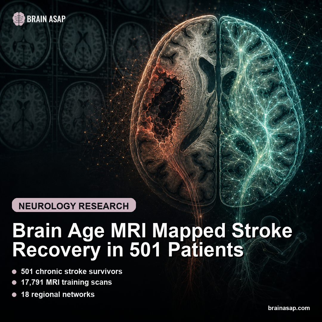
Brain Age MRI Mapped Stroke Recovery in 501 Patients
TL;DR: A Lancet Digital Health ENIGMA study found that larger stroke lesions made the damaged hemisphere look biologically older, while severe motor impairment was linked to younger-appearing contralesional networks, likely reflecting compensation.
Key Findings
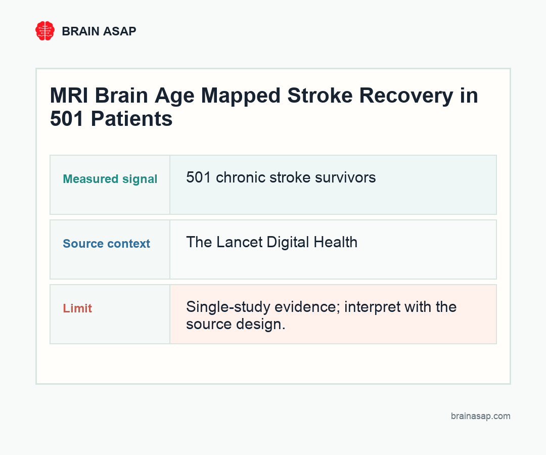
- 501 chronic stroke survivors: The ENIGMA Stroke Recovery Working Group dataset included participants from 34 cohorts in eight countries, all more than 180 days post-stroke.
- 17,791 MRI training scans: A UK Biobank sample trained the regional brain-age prediction model.
- 18 regional networks: Structural T1 MRI was used to estimate brain-predicted age difference across predefined functional subregions.
- Damaged side aged upward: Larger lesions were associated with higher ipsilesional regional brain age across multiple regions (β = 0.5420 to 0.9458).
- Opposite side looked younger: Larger lesions were linked to lower contralesional ventral attention/language brain age (β = -0.3747; 95% CI, -0.6961 to -0.0534).
Source: The Lancet Digital Health (2026) | Park et al.
Stroke recovery is not just an account of the tissue that was lost. It is also an account of how the remaining brain reorganizes around the damage. This study used deep learning-derived regional brain age to make that reorganization visible across 501 chronic stroke survivors.
Brain Age Made Stroke Damage Regional
Brain-predicted age difference, often shortened to brain-PAD, compares how old a brain region looks on MRI with the person’s actual chronological age. A positive value suggests older-appearing tissue; a negative value suggests younger-appearing tissue.
The study used a graph convolutional network trained on 17,791 UK Biobank scans, then applied it to 501 chronic unilateral stroke survivors. Instead of estimating one global brain age, the model examined 18 functional subregions.
The regional approach is the key. Stroke is focal, recovery is networked, and a single whole-brain number can blur motor-recovery biology.
The Damaged Hemisphere Looked Older
Larger total lesion size was associated with higher regional brain age in the ipsilesional hemisphere, meaning the side with the stroke looked biologically older across many regions. The reported beta range across significantly associated regions was 0.5420 to 0.9458.
That fits intuition. A large lesion disrupts tissue, connectivity, and downstream network health. The MRI model appears to capture that disruption as accelerated regional aging.
But the paper becomes more interesting when it looks at the other hemisphere. Stroke recovery is often about what undamaged circuits can still do.
The Contralesional Signal Ran in the Opposite Direction
Larger lesions were associated with lower brain-PAD in a contralesional ventral attention and language network region. In other words, part of the opposite hemisphere looked younger relative to age.
The authors interpret this as a compensation signal. More severe motor impairment was also linked to younger contralesional brain age in structural equation modeling, suggesting that the brain is reorganizing undamaged networks when the injured motor system cannot carry the load.
That is a subtle result. Younger-appearing tissue is not automatically better recovery. In this context, it likely reflects the brain’s attempt to adapt to severe damage, not proof that function has normalized.
Corticospinal Tract and Salience Network Led Motor Prediction
The machine-learning models identified three top predictors of motor outcomes: corticospinal tract lesion load, salience network lesion load, and contralesional frontoparietal brain-PAD.
The corticospinal tract result is expected because it is the major descending motor pathway. The salience network result is more network-level: it points to attention, relevance detection, and control systems that may influence how motor recovery unfolds.
The third predictor, contralesional frontoparietal brain age, is the most conceptually fresh. It suggests that MRI-derived regional age captures compensatory neuroplasticity that conventional lesion maps miss.
- Corticospinal tract: damage to the main motor pathway remained a strong predictor of worse motor outcome.
- Salience network: lesion load in attention and control circuitry added a network-level signal.
- Contralesional frontoparietal age: younger-appearing tissue on the opposite side marked compensatory recruitment rather than simple recovery.
A Biomarker for Rehabilitation Targeting Is Still Early
The study was retrospective and observational. It cannot prove that a younger-looking contralesional region causes compensation, nor can it say which rehabilitation strategy should be assigned to a specific patient based on regional brain age.
It also depends on harmonizing MRI data across 34 cohorts, which is both a strength and a headache. Multicohort diversity makes the result more robust, but differences in scanners, protocols, lesion segmentation, and clinical scoring can add noise.
Stroke rehabilitation needs biomarkers that capture both injury and adaptation. A regional brain-age map can help identify not only how much damage occurred, but which surviving networks are trying to reorganize around it.
AI-derived brain age is not just a mortality-clock gimmick here. It becomes a way to see the push-pull between damaged tissue and compensatory networks after stroke.
Why Younger-Looking Tissue Can Mean Strain, Not Success
Brain-age language can be misleading because “younger” sounds automatically good. In this study, the younger-appearing contralesional signal was associated with worse motor impairment, which changes the interpretation. It may be a marker of compensatory effort rather than a simple sign of healthier tissue.
The clinical interpretation is more complicated than “younger is better.” Compensation can be adaptive, inefficient, temporary, or even maladaptive depending on timing and circuit. A biomarker that captures compensatory recruitment can help identify which patients need therapies aimed at strengthening spared networks versus restoring activity in damaged pathways.
The frontoparietal and attention-network signals are especially relevant because motor recovery is not only about muscle commands. Planning, attention, error monitoring, and task selection all shape how movement is relearned after stroke.
Regional Brain Age Could Pair With Lesion Maps
Traditional lesion mapping is still essential. The corticospinal tract result in this study confirms that damage to the descending motor pathway remains one of the clearest predictors of motor outcome. Brain-age modeling should not replace that information.
The more realistic future is combination. A rehabilitation team can eventually look at lesion load, tract disruption, regional brain age, functional connectivity, and clinical performance together. That kind of multimodal profile would support more targeted therapy selection than lesion size alone.
For example, 2 patients can have similar lesion volumes but different recovery biology: one may have severe corticospinal disruption with little compensatory network signal, while another may show active contralesional recruitment that therapy could try to harness.
This paper does not deliver that clinical tool yet. It gives a reason to build it.
ENIGMA Scale Made the Stroke Recovery Signal More Credible
Single-center stroke MRI studies can be hard to generalize because scanner protocols, lesion-segmentation methods, rehabilitation settings, and outcome scoring can differ from one hospital to another. This paper gains force from ENIGMA scale: 34 cohorts across eight countries, with a large independent UK Biobank set used for training the age model.
That does not remove all harmonization problems, but it makes the result less likely to be a quirk of one scanner or one rehabilitation center. For a biomarker that might eventually guide recovery planning, the broader footprint is important.
The next question is whether regional brain-age patterns can predict who improves with specific therapy types. That would move the method from explaining recovery after the fact toward helping clinicians choose rehabilitation strategies while there is still time to shape plasticity.
Paper: Associations between contralesional neuroplasticity and motor impairment through deep learning-derived MRI regional brain age in chronic stroke (ENIGMA): a multicohort, retrospective, observational study. The Lancet Digital Health. 2026. DOI: 10.1016/j.landig.2025.100942
Authors: Park et al.
Study Design: Multicohort retrospective observational MRI study using a deep learning regional brain-age model.
Sample Size: 501 chronic unilateral stroke survivors from 34 cohorts in eight countries; 17,791 UK Biobank scans for model training.
Key Statistic: Higher corticospinal tract lesion load predicted poorer motor outcomes (β = -0.355; 95% CI, -0.446 to -0.267; P<0.0001), which were linked to younger contralesional brain age.