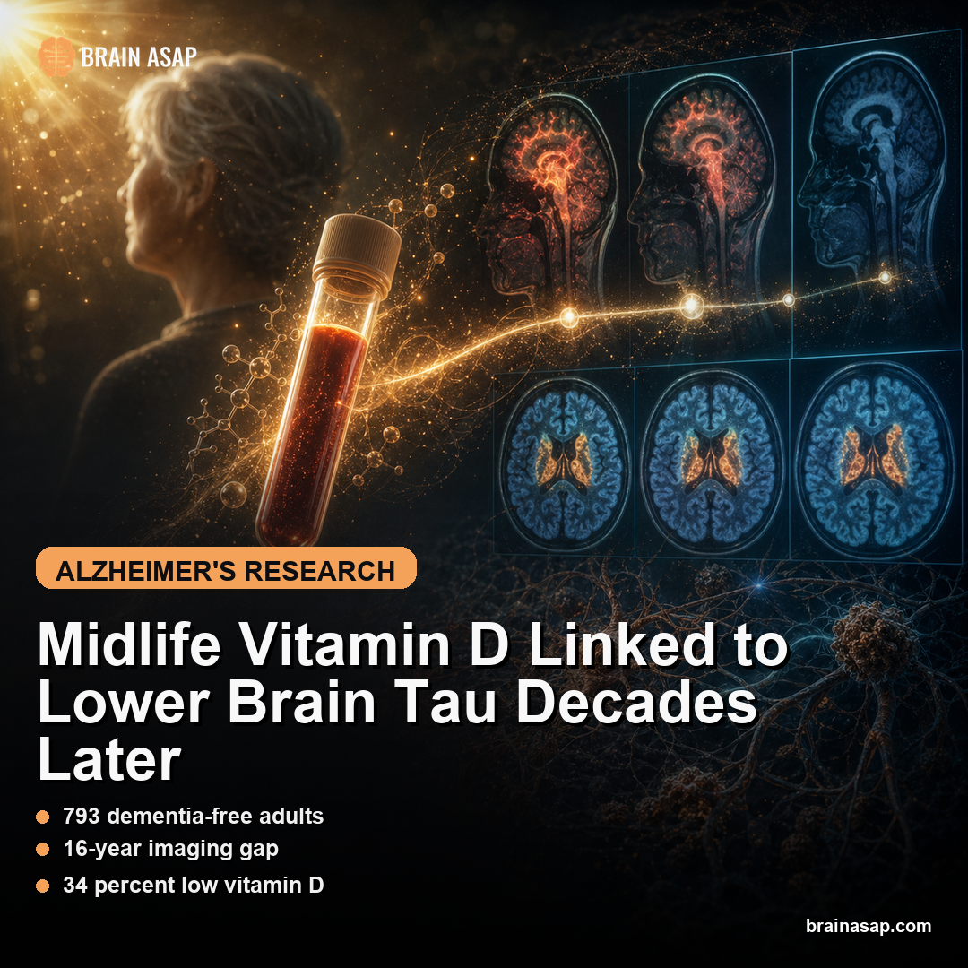
TL;DR: A 2026 study in Neurology Open Access found that in 793 dementia-free adults, higher vitamin D levels around age 39 were associated with less tau readout on PET brain imaging about 16 years later, while amyloid beta did not show the same relationship.
Key Findings
- 793 dementia-free adults: Participants had vitamin D measured in early midlife, at an average age of 39.
- 16-year imaging gap: About 16 years later, participants underwent positron emission tomography (PET), an imaging method that uses radioactive tracers to map biology in living tissue scans measuring tau and/or amyloid beta.
- 34% low vitamin D: A level below 30 ng/mL was classified as low, and about one-third of the cohort fell below that threshold.
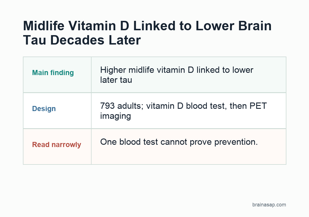
- Tau, not amyloid: Higher midlife vitamin D was associated with lower later tau readout on PET brain imaging, but not with amyloid beta burden.
- Single blood draw limit: Vitamin D was measured once, so the study cannot tell whether long-term vitamin D status or supplementation changed brain outcomes.
Source: Neurology Open Access (2026) | Mulligan et al.
Vitamin D is usually framed as a bone, immune, or general health nutrient.
Mulligan and other researchers tested a narrower brain-aging test: whether a blood vitamin D level measured around age 39 was associated with later PET imaging markers of Alzheimer’s-related proteins.
The association was specific. Higher midlife vitamin D tracked with lower later tau burden, while amyloid beta, another Alzheimer’s-related protein, did not show the same relationship.
The Study Looked Before Dementia, Not After
The strongest feature of the paper is timing.
Study details:
- 793 dementia-free adults: Participants had vitamin D measured in early midlife, at an average age of 39
- 16-year imaging gap: About 16 years later, participants underwent positron emission tomography (PET), an imaging method that uses radioactive tracers to map biology in living tissue scans measuring tau and/or amyloid beta
- 34% low vitamin D: A level below 30 ng/mL was classified as low, and about one-third of the cohort fell below that threshold
- Tau, not amyloid: Higher midlife vitamin D was associated with lower later tau readout on PET brain imaging, but not with amyloid beta burden
Participants were dementia-free when their vitamin D level was measured, and they were relatively young for an Alzheimer’s biomarker study: the average age was 39.
Many dementia-risk studies begin after brain pathology can already be well underway. This analysis asked whether a modifiable midlife factor was associated with later PET markers before dementia was present.
The cohort was followed for about 16 years before later brain imaging. That gives the analysis enough time to ask a real longitudinal test, even though it remains observational.
Higher Vitamin D Tracked With Lower Tau
Tau is one of the major proteins involved in Alzheimer’s disease. Amyloid beta tends to appear earlier in many models, while tau burden often tracks more closely with neurodegeneration and cognitive symptoms.
In this study, higher midlife vitamin D was associated with less later tau readout on PET brain imaging.
The scanned source material reports lower global and composite tau measures among people with vitamin D at or above 30 ng/mL, with beta estimates around -0.022 to -0.023 in secondary reporting.
The key point is direction and specificity: the association appeared for tau, not amyloid beta.
That finding is more informative than a generic “vitamin D is good for the brain” headline because tau and amyloid represent different parts of Alzheimer’s biology.
Tau Changed While Amyloid Did Not
The absence of an amyloid-beta association should keep expectations grounded.
If vitamin D were a broad Alzheimer’s shield in this dataset, one might expect a clearer readout across multiple biomarker systems.
Instead, the study points to tau.
That does not weaken the paper; it sharpens it.
Tau and amyloid are different biological processes, and risk factors do not necessarily move them in the same way.
A nutrient, hormone, vascular factor, or inflammatory state could plausibly relate to one pathway more than another.
- Tau readout: higher midlife vitamin D tracked with less later tau readout on PET brain imaging.
- Amyloid boundary: amyloid beta burden did not show the same relationship.
- Clinical limit: the result does not prove supplementation prevents dementia.
The clinical interpretation is cautious: this is not evidence that taking vitamin D supplements will prevent dementia.
It is evidence that vitamin D status in midlife deserves more attention in studies of tau biology.
30 ng/mL Was the Vitamin D Threshold
The study classified vitamin D above 30 ng/mL as high and values below that as low.
About 34% of participants had low vitamin D, while only 5% reported using vitamin D supplements.
Because supplement use was uncommon, the study was mostly observing variation in vitamin D status rather than a cohort dominated by supplementation.
Still, vitamin D is entangled with sun exposure, diet, health behavior, adiposity, geography, and medical care.
The authors adjusted for factors such as age, sex, and depressive symptoms, but residual confounding is always possible.
People with higher vitamin D may differ from those with lower vitamin D in ways that are hard to fully measure.
One Blood Test Cannot Show Dementia Prevention
The biggest limitation is that vitamin D was measured once.
A single blood draw around age 39 does not necessarily represent a person’s vitamin D exposure over the next decade and a half.
The study also did not randomize supplementation. That means it cannot answer the test most people would immediately ask: if my vitamin D is low, will correcting it reduce future tau burden?
The study points researchers toward a testable midlife target.
If later trials or repeated-measure cohorts show that sustained vitamin D sufficiency changes tau trajectories, the public-health implications could be substantial because vitamin D deficiency is common and modifiable.
The reasonable action is not to megadose vitamin D for dementia prevention.
It is to know whether vitamin D is low and correct deficiency through routine care when appropriate, because deficiency is common, measurable, and already relevant to bone and general health.
Why Tau Specificity Makes the Result More informative
If the result had shown a vague association with every Alzheimer’s biomarker, it might have been easier to suspect a broad healthy-user effect.
The tau-specific pattern is more biologically intriguing.
It raises the possibility that vitamin D status can relate to neurodegenerative or inflammatory processes that are more tightly coupled to tau than to amyloid deposition.
Vitamin D receptors are present in brain-relevant tissues, and vitamin D has been studied in immune regulation, vascular biology, oxidative stress, and neuroinflammation.
None of that proves causality here, but it offers plausible pathways worth testing.
The study also lands in a prevention-relevant life-stage window.
Early midlife is late enough that long-term lifestyle and medical factors are measurable, but early enough that dementia is usually still far in the future.
That is why the average baseline age of 39 is not a side detail; it changes what the result can mean.
What a Supplement Trial Would Need to Prove
A future trial would need to do more than raise blood vitamin D.
It would need to track whether sustained correction of low vitamin D changes tau readouts on PET brain imaging, cognitive outcomes, or both, ideally while accounting for baseline deficiency, sex, ancestry, kidney function, body composition, sunlight exposure, diet, and APOE genotype.
A trial like that would ask a harder test than whether low vitamin D is associated with later tau.
The current study helps justify the work because vitamin D is cheap to measure, deficiency is modifiable, and the reported readout was tied to a specific Alzheimer’s-related biomarker rather than a vague brain-health score.
The Dementia-Free Sample Changes the Stakes
Because participants were dementia-free when vitamin D was measured, the finding is about early risk biology rather than treatment after symptoms appear.
A midlife blood level associated with later tau is more credible as a consequence of dementia-related frailty, reduced outdoor activity, or advanced illness.
Measuring vitamin D around age 39 does not eliminate reverse causation or confounding.
It does make the timing more informative than a study that measures vitamin D only after older adults have already developed major cognitive decline.
Citation: DOI: 10.1212/WN9.0000000000000057. Mulligan et al. Association of Circulating Vitamin D in Midlife With Increased Tau-PET Burden in Dementia-Free Adults. Neurology Open Access. 2026;2(2):e000057
Study Design: Longitudinal observational biomarker study with midlife serum vitamin D and later PET imaging.
Sample/Model: 793 dementia-free adults at baseline; amyloid and/or tau PET imaging about 16 years later.
Key Statistic: Higher vitamin D status was associated with less later tau readout on PET brain imaging, while amyloid beta burden showed no association.
Caveat: Single-study evidence; interpret with the source design and sample.