TL;DR: A 2026 study in The British Journal of Psychiatry found that in first-episode psychosis, MRI deviations from age-matched brain norms at illness onset helped predict which patients would carry heavier symptom burdens and poorer functioning for years afterward.
Key Findings
- 100,000-scan reference model: Researchers used a lifespan normative MRI model built on more than 100,000 scans to measure how far each patient’s grey matter volume diverged from age- and sex-expected values at first-episode psychosis.
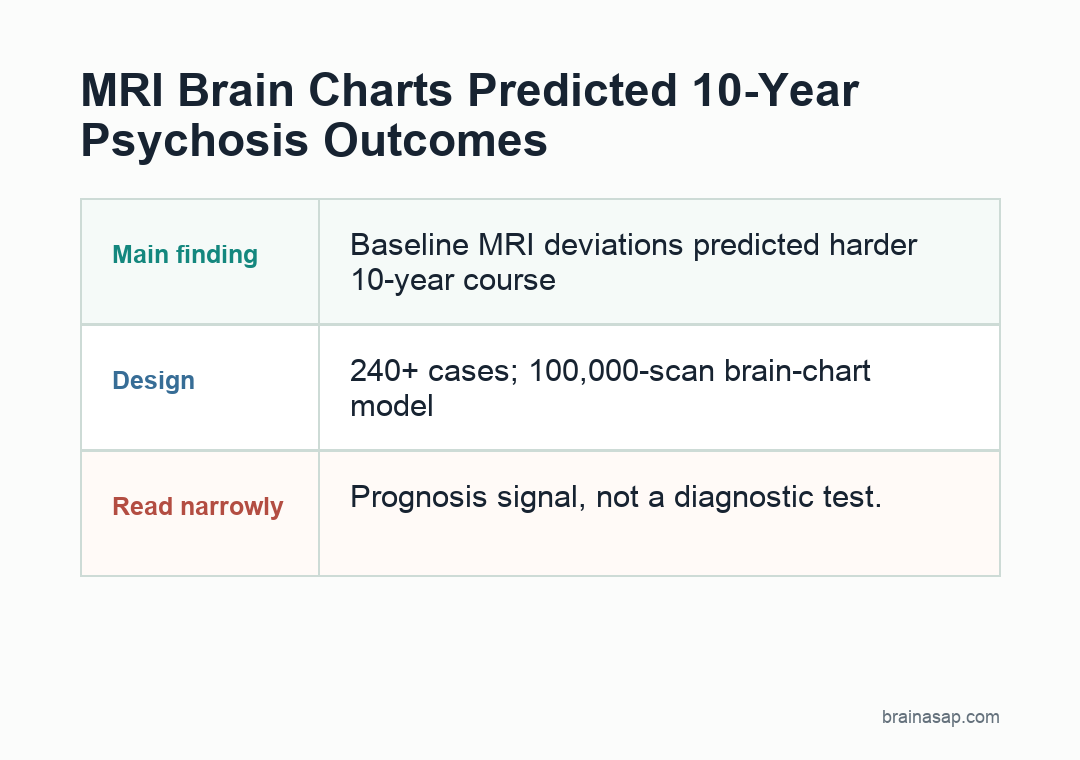
- More than 240 early psychosis cases: Baseline MRI from a Spanish first-episode psychosis cohort was linked to clinical follow-up at 1, 3 and roughly 10 years, giving the study a much longer horizon than most imaging papers manage.
- Left superior temporal gyrus readout: More negative baseline deviations in the left superior temporal gyrus tracked steeper long-term worsening in positive symptoms, negative symptoms and overall functioning.
- Broca’s area also mattered: Reduced grey matter volume in Broca’s area at illness onset predicted a tougher clinical trajectory, suggesting language-related frontal cortex may be part of the early risk profile.
- Function, not just symptoms: The imaging deviations were linked to symptom scales and to Global Assessment of Functioning, supporting individualized MRI charts over broad case-control averages.
Source: The British Journal of Psychiatry (2026) | Muñoz-Caracuel et al.
Psychosis care still starts with a blunt problem: 2 patients can look similar in the clinic on day one and then diverge dramatically over the next decade.
This paper tries to get ahead of that split by asking whether the brain’s starting point, measured against a huge lifespan MRI reference set, can tell us who is more likely to struggle long term.
Why a 100,000-Scan Brain Chart Could Matter More Than Another Group Average
Psychosis imaging has had a long credibility problem.
Study details:
- 100,000-scan reference model: Researchers used a lifespan normative MRI model built on more than 100,000 scans to measure how far each patient's grey matter volume diverged from age- and sex-expected values at first-episode psychosis
- More than 240 early psychosis cases: Baseline MRI from a Spanish first-episode psychosis cohort was linked to clinical follow-up at 1, 3 and roughly 10 years, giving the study a much longer horizon than most imaging papers manage
- Left superior temporal gyrus readout: More negative baseline deviations in the left superior temporal gyrus tracked steeper long-term worsening in positive symptoms, negative symptoms and overall functioning
- Broca's area also mattered: Reduced grey matter volume in Broca's area at illness onset predicted a tougher clinical trajectory, suggesting language-related frontal cortex may be part of the early risk profile
Group studies repeatedly show that patients, on average, differ from controls in certain brain regions, yet those differences rarely translate into a tool a psychiatrist could use when a single patient shows up in crisis.
This paper takes aim at that gap by replacing the usual case-control logic with a normative model: instead of asking whether psychosis brains differ on average, it asks whether an individual patient’s brain sits outside the range expected for their age and sex.
That may seem like a subtle shift, but it changes the clinical test. Averages are helpful for papers; departures from a personal reference range are more helpful for prognosis.
To do that, the authors leaned on one of the largest brain-chart efforts available, a lifespan MRI reference dataset with more than 100,000 scans.
Each patient entering first-episode psychosis could then be mapped onto that chart and assigned regional deviations in cortical and subcortical grey matter volume.
The study cohort itself also deserves attention.
These were drug-naive or minimally treated patients recruited through Spain’s PAFIP early psychosis program, with baseline MRI near illness onset and repeated follow-up across years rather than weeks.
The prognosis setup had three important pieces:
- Early scan: MRI was collected near the first episode, before years of illness could blur the readout.
- Normative comparison: each brain was compared with age- and sex-expected volume from a large reference model.
- Long follow-up: symptoms and functioning were tracked across years, not just weeks.
Prognostic tools only earn their keep if they tell us something about the future, not just the acute episode that is already in front of us.
In practice, the authors were not trying to find one definitive “psychosis lesion.” They were testing whether the brain’s starting geometry could nudge long-term trajectories toward worse or better outcomes.
That is a more realistic ambition, and it fits what clinicians actually need: triage, not certainty.
How Left Superior Temporal Gyrus Deviations Forecast a Harder Road
The strongest anatomical readout emerged in the left superior temporal gyrus, a region often implicated in language processing, auditory integration and the broader cortical machinery that helps organize meaning.
Patients whose baseline grey matter volume in that region fell further below normative expectations went on to show more severe trajectories across positive symptoms, negative symptoms and functioning.
That finding is notable because it bridges several domains that are usually split apart in psychosis research.
Positive symptoms and functional decline are often analyzed as separate outcomes, while negative symptoms get treated as a partly independent syndrome.
Here, one regional deviation touched all three.
That does not show the superior temporal gyrus single-handedly causes psychosis progression.
It means its early structural departure can act as a compact readout of deeper developmental vulnerability.
The superior temporal cortex has long been linked to auditory hallucinations and disorganized language processing, but the paper’s payoff is broader than symptom mechanism.
A patient whose baseline MRI already shows a bigger drop relative to expected developmental norms is starting the illness from a more fragile cortical architecture.
That, in turn, can make recovery slower, relapse more likely, or psychosocial repair harder to sustain even if acute psychosis is brought under control.
That is where normative modeling helps.
A raw grey matter value can be hard to interpret across ages and sexes.
A deviation from expected volume is easier to frame clinically: this patient’s cortex is not just small or large; it is further from the normal developmental lane than it should be.
Broca’s Area Kept Showing Up in Long-Term Symptom Trajectories
The other region the paper keeps pulling into focus is Broca’s area, classically associated with speech production but functionally connected to much more than talking.
In psychosis, it sits near the crossroads of inner speech, cognitive control and language organization, all processes that can go sideways early in schizophrenia-spectrum illness.
When the authors modeled trajectories over time, more negative baseline deviations in Broca’s area were linked to more severe symptom progression and weaker functional recovery.
That is an important clue because it points away from the idea that poor outcome is explained purely by subcortical dopamine burden or by social circumstances after diagnosis.
The frontal language-control system appears to carry part of the prognostic readout too.
There is also something clinically intuitive here.
Patients who struggle early with language organization, thought flow and executive control often have a harder time navigating school, work and relationships even when florid psychosis settles down.
A structural marker in Broca’s territory fits that lived pattern. It suggests the MRI is capturing a brain system tied to both symptom expression and real-world adaptation.
The study does not claim that these regions are sufficient for prediction on their own.
But it does suggest that the most informative anatomy does not have to be the most dramatic-looking abnormality.
Small, person-specific deviations in regions tied to language and social cognition can carry more prognostic value than a blunt “whole brain volume loss” explanation.
What This Adds to the First-Episode Psychosis Playbook
A lot of prognostic work in psychosis depends on variables clinicians already know how to collect: duration of untreated psychosis, early symptom severity, family support, substance use, medication response. Those matter.
But they still do not fully explain why some patients stabilize while others accumulate disability.
This paper suggests that a baseline MRI, interpreted through a developmental reference model, can add a helpful biological layer.
The biggest practical implication is not “scan everyone and let the computer decide.” It is more targeted.
If a first-episode service can identify patients whose baseline cortical deviations point toward a riskier long-term course, those patients justify closer monitoring, more aggressive relapse prevention, family-focused care and vocational support before decline becomes entrenched.
That is where function becomes especially important. Symptom improvement is not the same thing as life recovery.
Someone can have fewer delusions and still lose educational momentum, social confidence or occupational footing.
By showing links to the Global Assessment of Functioning, the study nudges imaging biomarkers closer to the outcomes patients actually care about.
It also reframes MRI from a diagnostic tool to a developmental context tool. The scan is not being used to confirm psychosis.
It is being used to estimate how far a patient’s brain architecture has drifted from the developmental path one would expect. That is a more plausible clinical role for imaging in psychiatry.
This Still Is Not a Ready-to-Deploy Prognosis Machine
The paper is promising, but it is not a turnkey prediction engine.
The cohort comes from a single early-intervention program in Spain, even if the follow-up is impressively long.
Replication across other health systems, scanners and ethnic backgrounds still matters if this is going to become clinically robust rather than academically informative.
There is also the usual MRI biomarker challenge: an association with outcome is not automatically a decision rule.
To become truly helpful, the normative deviations would need to improve prediction beyond standard clinical variables in a way that changes management.
The paper gets us closer to that test, but it does not fully answer it.
Another caution is that psychosis is not one disease with one biological script. The point of normative modeling is partly to respect that heterogeneity.
So the right way to read this study is not that psychosis “lives” in the superior temporal gyrus or Broca’s area.
It is that early divergence in these regions can act as a marker of who may need more support over time.
Still, this is one of the more believable routes MRI has offered toward psychiatric usefulness.
Instead of claiming to diagnose schizophrenia from a scan, the authors ask a narrower, more defensible test: can the brain’s distance from normal development at first episode tell us something about where a patient is heading? On the evidence here, the answer looks increasingly like yes.
Citation: DOI: 10.1192/bjp.2025.105. Muñoz-Caracuel et al. Predicting clinical and functional trajectories in individuals with first-episode psychosis by baseline deviations in grey matter volume
The British Journal of Psychiatry.
2026;228(5):419-427..
Study Design: Cohort study
Sample/Model: 100,000-scan reference model: Researchers used a lifespan normative MRI model built on more than 100,000 scans to measure how far each patient’s grey matter volume diverged from age- and sex-expected values at first-episode psychosis.
Key Statistic: More than 240 early psychosis cases: Baseline MRI from a Spanish first-episode psychosis cohort was linked to clinical follow-up at 1, 3 and roughly 10 years, giving the study a much longer horizon than most imaging papers manage.
Caveat: Single-study evidence; interpret with the source design and sample.