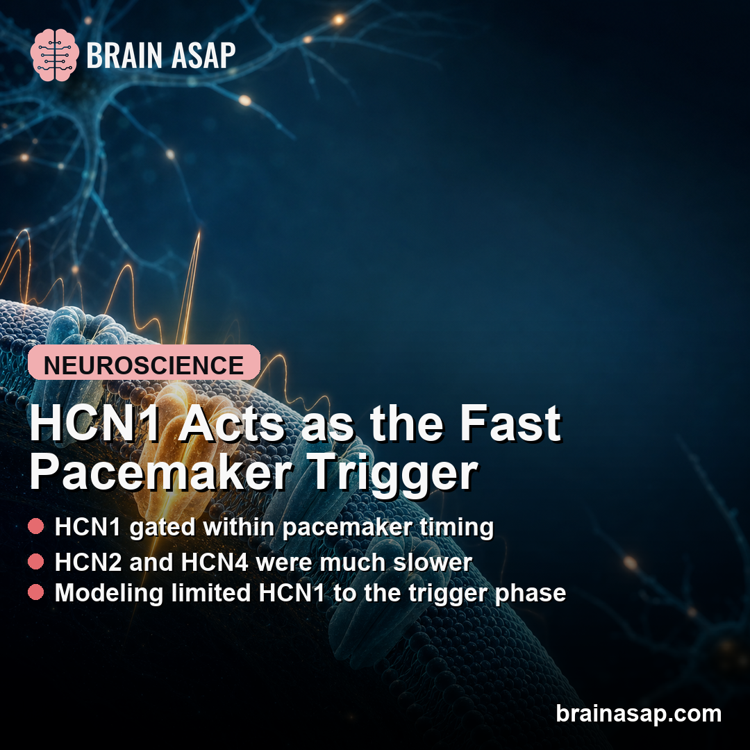
TL;DR: A 2026 Nature Communications study found that HCN1, an ion channel that helps neurons start rhythmic electrical firing, was fast enough to act as the trigger for neuronal pacemaker depolarization, while HCN2 and HCN4 mainly appeared too slow to drive each action-potential cycle.
Key Findings
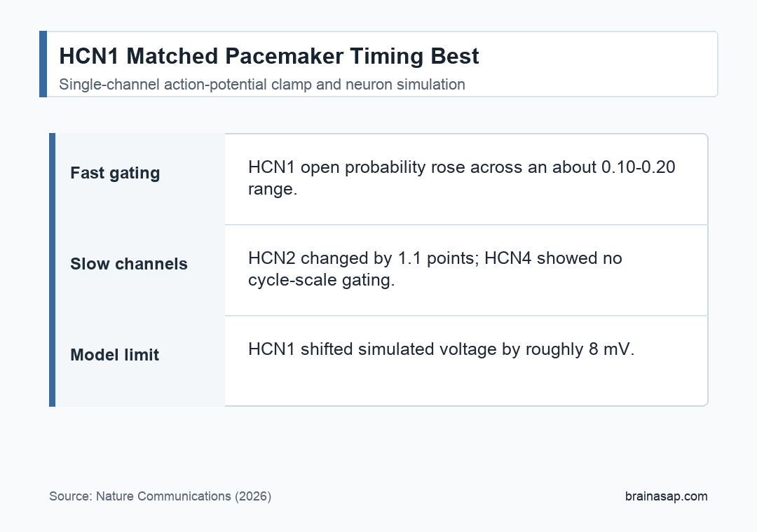
- HCN1 was the fast channel: In slow action-potential clamp experiments, HCN1 open probability increased from about 0.10 to about 0.20 during pacemaker depolarization, a roughly 96% increase.
- HCN2 barely moved: HCN2 showed only a 1.1 percentage-point increase in open probability at 2.77 Hz, on top of a background open probability near 20.5%.
- HCN4 stayed too slow: HCN4-like channels showed no meaningful time-dependent gating during the same pacemaker depolarization window.
- HCN1 relaxed much faster: Researchers estimated HCN1 activation during pacemaker depolarization at 43 plus or minus 11 ms, while HCN2 and HCN4 recovery processes were in the 8 to 15 second range.
- The model limited HCN1’s role: Simulations suggested HCN1 alone depolarized the model neuron only from -81 mV to about -73 mV, roughly the first quarter of the ramp toward threshold.
Source: Nature Communications (2026) | Enke et al.
HCN1 Was Fast Enough for Pacemaker Timing
HCN channels, short for hyperpolarization-activated cyclic nucleotide-modulated channels, help shape rhythmic electrical activity in neurons. These channels are often discussed as pacemaker channels because they can carry inward current during the quiet phase between action potentials.
The problem is timing. Neuronal pacemaker firing can occur at several hertz or faster, while older voltage-clamp measurements suggested that many HCN channels open and close too slowly to explain each firing cycle.
Researchers tested three mouse channel types: HCN1, HCN2, and an HCN4-like mutant called HCN4V487E. The HCN4 mutant was used because its single-channel current was easier to resolve while retaining HCN4-like gating behavior.
- HCN1: The fastest isoform, tested for whether it could change state during the pacemaker depolarization itself.
- HCN2: A slower channel with a measurable open probability but little cycle-by-cycle movement.
- HCN4-like channel: A slow channel that mainly helped define the comparison against HCN1.
Action-Potential Clamp Tracked Single Channels During Firing
The central method was a dynamic action-potential clamp, a recording approach that plays a neuron-like voltage waveform back to a cell while measuring channel current. Instead of asking how a channel behaves under a square pulse, researchers measured channel opening during a pacemaker-shaped voltage trajectory.
The experiments used Xenopus oocytes, a standard expression system for ion-channel studies, because the method required extremely stable patches and femtosiemens-scale resolution. That resolution was needed because individual HCN channels conduct very tiny currents.
Researchers ran two main firing patterns:
- Fast 10 Hz trains: These tested whether channels could keep up with a rapid pacemaker rhythm.
- Slower 2.77 Hz trains: These gave slower channels more time to reveal any activation or deactivation during the pacemaker ramp.
- Repeated action potentials: At least 50 action potentials were played in a train after a brief deactivation pulse.
HCN2 and HCN4 Behaved as Background Conductances
HCN2 and HCN4 were not inactive. Both produced meaningful open probability during pacemaker depolarization. The important result was that their open probability stayed mostly flat within the cycle.
At 10 Hz, HCN2 and HCN4-like channels showed open channels, but the measured open probability did not rise and fall in the way expected for a channel directly timing each pacemaker depolarization. Their conductance recovered over many seconds instead.
At 2.77 Hz, HCN2 showed a small detectable change: a 1.1 percentage-point increase on top of a background open probability near 20.5%. Researchers interpreted that as minimal contribution to cycle-by-cycle pacemaker gating.
HCN4-like channels showed even less cycle-scale gating. The study argues that HCN2 and HCN4 may still matter by setting a neuron’s electrical working point, especially because these channels are more sensitive to cyclic AMP, a signaling molecule that can tune pacemaker activity.
HCN1 Acted Like an Initial Trigger, Not the Whole Engine
HCN1 behaved differently. During the slower action-potential waveform, HCN1 open probability increased from about 0.10 to 0.20 during pacemaker depolarization. That was a roughly 96% increase in open probability over the relevant window.
The timing also separated HCN1 from the other channels. Researchers estimated HCN1 activation during pacemaker depolarization at 43 plus or minus 11 ms.
By contrast, HCN2 and HCN4 recovery processes were estimated in the 8 to 15 second range.
The modeling result kept the conclusion from becoming too broad. In a simplified suprachiasmatic-nucleus neuron model, HCN1 alone moved the membrane voltage from -81 mV to about -73 mV.
That covered only the initial part of the route toward firing threshold.
- Trigger phase: HCN1 can start the pacemaker depolarization from a very negative membrane voltage.
- Takeover phase: Other depolarizing currents, such as T-type calcium channels, likely need to continue the ramp toward threshold.
- Tuning phase: HCN2, HCN4, and cyclic AMP sensitivity may tune the background voltage and responsiveness of the system.
The Caveat Is Translation From Oocytes to Living Neurons
This was a detailed biophysical study, not a direct recording from an intact brain circuit. The channels were expressed in Xenopus oocytes, and the recordings were done at room temperature because the required patch stability did not tolerate higher temperatures.
Living neurons contain mixtures of channel types, accessory proteins, heteromeric channels, and other conductances. Temperature also changes channel kinetics, so the oocyte recordings should be read as channel-timing evidence rather than a complete brain-circuit model.
The study authors argue that the basic timing pattern remains informative, but the exact numbers should not be treated as a direct measurement of every pacemaker neuron in the brain.
The result supports a narrow interpretation: HCN1 is the best candidate among the tested HCN channels for the fast trigger component of neuronal pacemaking. HCN2 and HCN4 may still shape excitability, but the data do not support them as the main cycle-by-cycle drivers in the tested conditions.
Citation: DOI: 10.1038/s41467-026-72257-3. Enke et al. HCN1 is a primary HCN Pacemaker Channel in Neurons. Nature Communications. 2026;17:3745.
Study Design: Single-channel electrophysiology using dynamic action-potential clamp plus a simplified neuron simulation.
Sample/Model: Mouse HCN1, HCN2, and HCN4-like channels expressed in Xenopus oocytes, with simulated suprachiasmatic-nucleus neuron dynamics.
Key Statistic: HCN1 open probability increased by about 96% during slow pacemaker depolarization, while HCN2 increased only 1.1 percentage points and HCN4-like channels showed no meaningful cycle-scale gating.
Caveat: The recordings used expressed channels at room temperature, so intact neurons may show additional effects from temperature, channel mixtures, and other conductances.