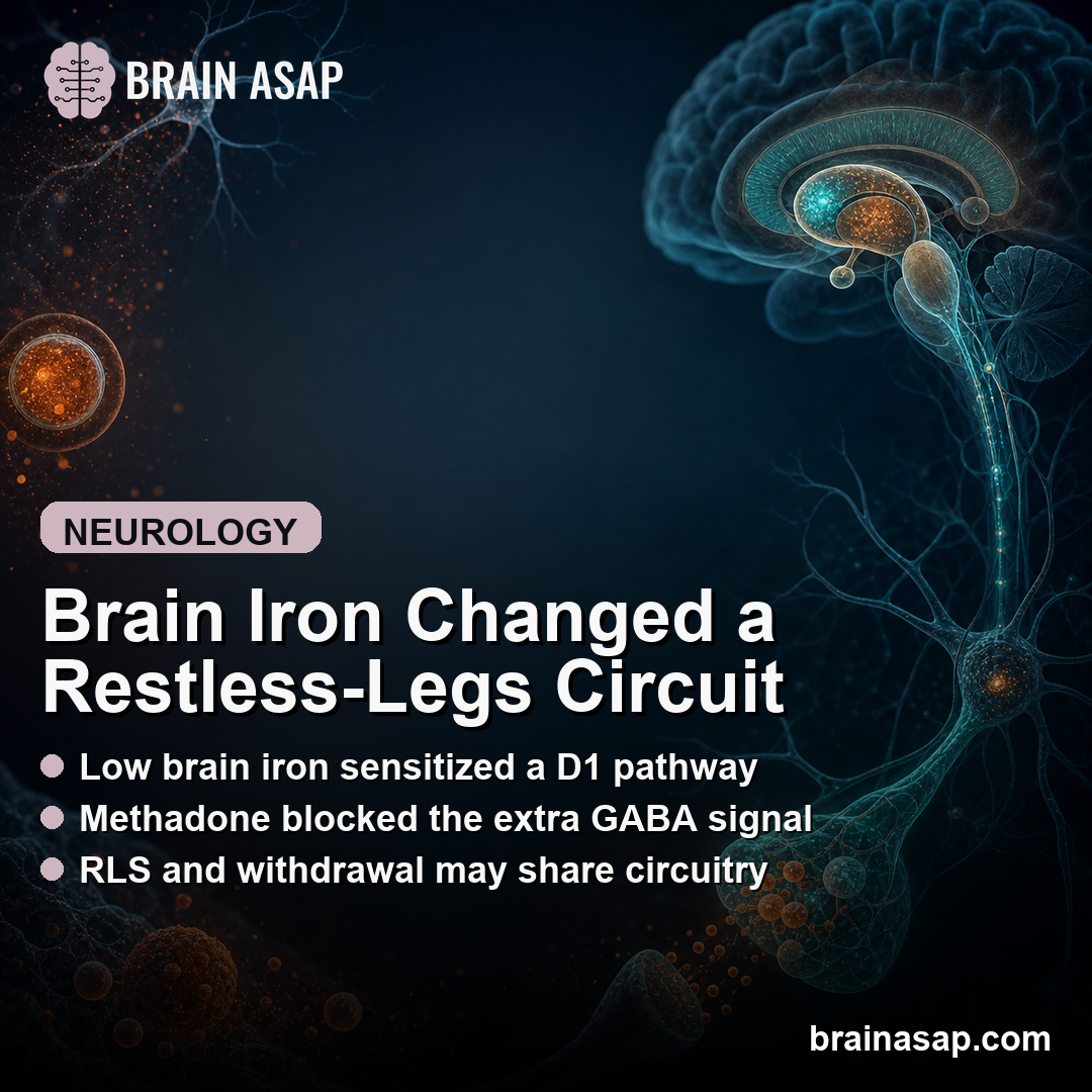
TL;DR: A 2026 mouse study in Neuropsychopharmacology found that brain iron deficiency made an opioid-responsive striatal pathway unusually sensitive to D1 dopamine receptor stimulation, pointing to a possible circuit mechanism for restless legs syndrome and opioid-withdrawal restlessness.
Key Findings
- Iron-deficient mouse model: Researchers used a low-iron diet to create brain iron deficiency (BID), a rodent model used to study restless legs syndrome biology.
- Low-dose dopamine test: The D1 receptor agonist SKF81297 at 1.5 mg/kg increased GABA release in the entopeduncular nucleus (EPN) in BID mice, but not in control-diet mice.
- Methadone-sensitive pathway: Methadone at 10 mg/kg reduced basal EPN GABA release and prevented the low-dose SKF81297-induced GABA increase in BID mice.
- Behavior split: BID did not increase D1-driven locomotor activation, suggesting the sensitized pathway was not the main movement-activation circuit.
- Receptor expression changed: BID reduced striatal mRNA expression for mu-opioid receptors and adenosine A1 receptors, while D1 receptor expression did not significantly change.
Source: Neuropsychopharmacology (2026) | Valle-Leon et al.
Brain Iron Deficiency Made One Restless-Legs Circuit More Dopamine-Sensitive
Restless legs syndrome (RLS) is marked by an urge to move that appears during rest, improves with movement, and often worsens at night.
Iron biology is part of that symptom pattern because altered brain iron homeostasis has been linked to RLS, even when ordinary blood measures do not fully explain the symptoms.
Researchers used a brain iron deficiency (BID) mouse model to test whether low brain iron changes dopamine and opioid signaling inside basal ganglia circuitry.
The focus was not simply whether mice moved more. Researchers asked which circuit responded differently when D1 dopamine receptors and mu-opioid receptors were pharmacologically stimulated.
- D1 dopamine receptors: These receptors respond to dopamine and can drive movement-related basal ganglia activity.
- Mu-opioid receptors: These receptors respond to opioid drugs and endogenous opioid signaling, and opioid medications can reduce severe RLS symptoms in selected patients.
- Entopeduncular nucleus: The EPN is the rodent counterpart of the internal globus pallidus, a basal ganglia output region involved in movement and motivational circuitry.
The main result was circuit-specific. A low dose of the D1 receptor agonist SKF81297 triggered EPN GABA release in BID mice.
The same dose did not produce that GABA response in control mice. In this EPN pathway, GABA release is treated as a readout of striatal neurons feeding into basal ganglia output.
In this model, low brain iron did not make every dopamine pathway stronger. It made one opioid-responsive striatal-EPN pathway react differently.
Methadone Blocked the Extra GABA Signal in Iron-Deficient Mice
The opioid arm of the experiment sharpened the interpretation. Researchers gave BID mice methadone at 10 mg/kg, then tested the same low SKF81297 dose.
Methadone reduced spontaneous EPN GABA release and prevented the SKF81297-induced increase.
The study was not a test of methadone as an RLS treatment in people. Instead, a mu-opioid receptor agonist changed the same circuit readout that became dopamine-sensitive under brain iron deficiency.
Researchers interpret the result as evidence that BID selectively increases sensitivity in striosomal MOR-D1R neurons, a striatal neuron population that carries both mu-opioid and D1 dopamine receptor signaling.
The evidence is still indirect because the study measured EPN GABA release after systemic drugs rather than directly recording every striosomal neuron involved.

Movement Activation Did Not Track the Same Circuit Response
The behavioral readout did not simply mirror the fiber-photometry response. SKF81297 increased locomotor activity in mice, but BID did not amplify that movement response.
In control-diet mice, researchers first tested SKF81297 dose response and estimated an EC50 of 2.5 mg/kg. They then compared low doses in control and iron-deficient mice.
The BID mice did not show stronger locomotor activation, and the lower dose response looked reduced rather than exaggerated.
The behavior-physiology split led to a circuit distinction:
- Matrix D1 neurons: These neurons appear to drive much of the D1 agonist locomotor activation.
- Striosomal MOR-D1 neurons: These neurons appear more relevant to the methadone-sensitive EPN GABA response.
- Different outputs: The study argues that movement activation and the opioid-responsive EPN pathway can diverge instead of reflecting one simple dopamine effect.
Reserpine experiments supported that separation. Reserpine depletes dopamine, allowing researchers to test postsynaptic D1 responses with less interference from endogenous dopamine.
In reserpinized mice, methadone no longer produced its strong locomotor effect and did not modify the SKF81297 response.
Lower Mu-Opioid and A1 Receptor Expression Fit the Sensitivity Model
The molecular readout also pointed toward altered modulation of D1 signaling. In striatal tissue from BID mice, researchers measured mRNA expression for Drd1, Oprm1, and Adora1.
Those genes encode D1 dopamine receptors, mu-opioid receptors, and adenosine A1 receptors.
D1 receptor expression did not significantly change. Mu-opioid receptor and A1 receptor expression were lower in the iron-deficient group.
Mu-opioid receptors and adenosine A1 receptors can modulate D1 signaling. Lower inhibitory or modulatory receptor expression could help explain why a low D1 agonist dose produced a stronger EPN GABA response in the BID condition.
- Drd1: D1 receptor expression stayed broadly comparable between groups.
- Oprm1: Mu-opioid receptor expression was significantly reduced in BID mice.
- Adora1: Adenosine A1 receptor expression was also significantly reduced in BID mice.
The receptor-expression result does not prove the whole circuit mechanism by itself. It gives a plausible molecular backdrop for the physiological result: the D1-sensitive EPN GABA response changed even without an obvious increase in D1 receptor expression.
The Human Claim Is Mechanistic, Not a Treatment Recommendation
The study connects two clinical observations: RLS often involves brain iron biology, and opioid withdrawal can produce RLS-like restlessness. If those states share an opioid-responsive striatal pathway, the overlap becomes more biologically specific.
The finding also gives a reason to be precise about opioid-related interpretations. Methadone blocked a circuit readout in mice.
The paper is not a human trial, not a dosing study, and not evidence that opioid treatment should be broadened. It is a mechanistic study of how low brain iron can alter dopamine-opioid circuit responsiveness.
Several limits keep the result bounded:
- Mouse model: Dietary BID is useful for RLS biology but does not reproduce every feature of human RLS.
- Systemic drugs: SKF81297 and methadone were administered systemically, so indirect effects from other brain regions cannot be fully excluded.
- Indirect neuron attribution: The striosomal MOR-D1R interpretation is based on circuit logic and prior work, not direct recording of every target neuron in this experiment.
The practical takeaway is narrower and stronger: brain iron deficiency may sensitize an opioid-responsive dopamine pathway that affects basal ganglia output. That mechanism could help explain why RLS involves both dopamine and opioid biology without reducing the condition to a generic movement problem.
Citation: DOI: 10.1038/s41386-026-02413-2. Valle-Leon et al. Dopaminergic hypersensitivity of the opioid-responsive striatal-entopeduncular pathway in a rodent model of restless legs syndrome. Neuropsychopharmacology. 2026.
Study Design: Mouse circuit study using a dietary brain iron deficiency model, locomotor testing, fiber photometry of EPN GABA release, and striatal receptor mRNA measurement.
Sample/Model: Male and female C57BL/6 mice, with postweaning low-iron diet exposure used to model brain iron deficiency relevant to restless legs syndrome.
Key Statistic: Low-dose SKF81297 at 1.5 mg/kg increased EPN GABA release in iron-deficient mice but not control mice; methadone at 10 mg/kg blocked that enhanced signal.
Caveat: The study is preclinical and uses systemic drug challenges, so it supports a circuit hypothesis rather than a direct treatment recommendation for patients.