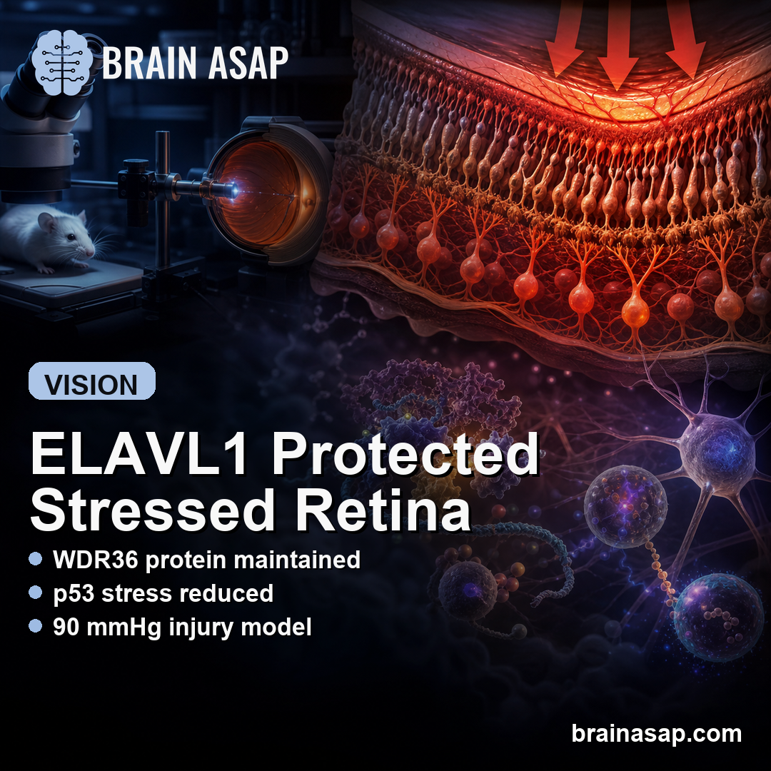
TL;DR: A 2026 study in Open Life Sciences reported that ELAVL1, an RNA-binding protein, protected stressed retinal cells by maintaining WDR36 protein, reducing p53 activation, and limiting calcium overload in cell and acute mouse pressure-injury models.
Key Findings
- OGD/R cell stress: Oxygen-glucose deprivation/reoxygenation reduced ELAVL1 and WDR36 protein in R28 retinal precursor cells.
- mRNA-protein mismatch: WDR36 protein fell under stress even though WDR36 mRNA did not, pointing to post-transcriptional regulation.
- ELAVL1 binding: RNA immunoprecipitation showed ELAVL1 enrichment of WDR36 mRNA, supporting direct binding between ELAVL1 and the WDR36 transcript.
- WDR36-dependent rescue: WDR36 knockdown reversed ELAVL1 overexpression benefits on cell viability, ATP, calcium overload, and p53/ER-stress markers.
- 90 mmHg mouse model: In acute intraocular-pressure elevation mice, ELAVL1 overexpression reduced retinal injury and apoptosis, but WDR36 knockdown weakened that protection.
Source: Open Life Sciences (2026) | Meng et al.
Retinal ganglion cell injury is central to glaucoma-related vision loss, but pressure is not the only biological problem. Stressed retinal cells also face mitochondrial dysfunction, calcium overload, p53 signaling, and endoplasmic-reticulum stress.
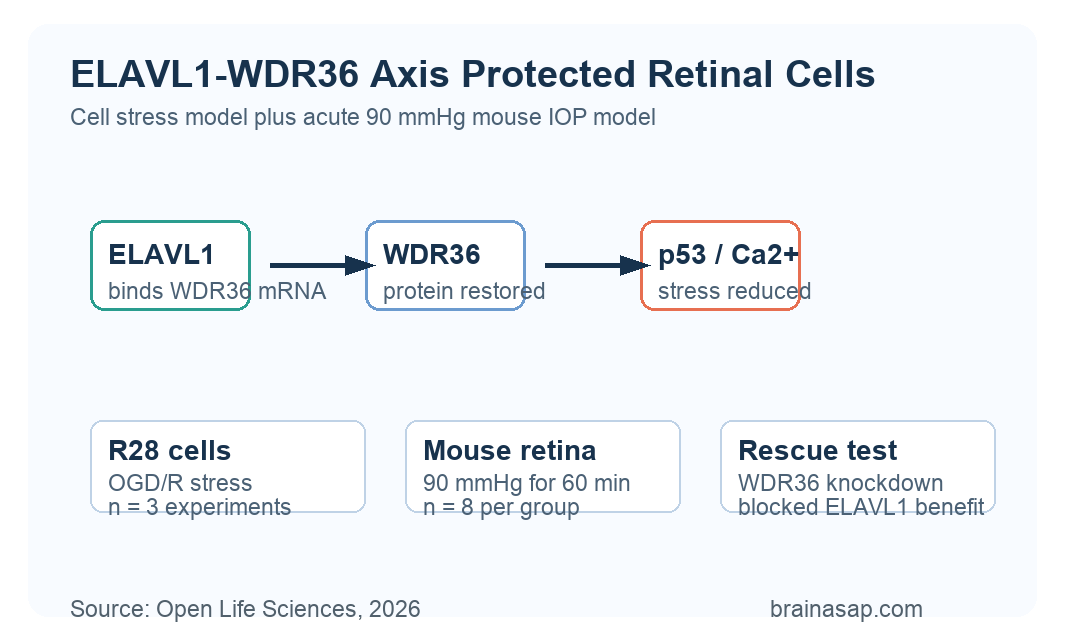
Meng et al. tested whether ELAVL1 helps retinal cells survive acute stress by regulating WDR36, a gene previously reported in glaucoma-related research.
The study used cell models and an acute mouse pressure-injury model, not patients with chronic glaucoma.
OGD/R Stress Lowered ELAVL1 and WDR36 Protein
The cell model used R28 retinal precursor cells exposed to oxygen-glucose deprivation/reoxygenation, or OGD/R. This model simulates ischemia-like cellular stress rather than the full complexity of human retinal disease.
Under OGD/R conditions, WDR36 protein decreased. WDR36 mRNA stayed relatively stable, which suggested that the problem was not simple loss of gene transcription.
- Cell viability: OGD/R reduced survival of R28 retinal precursor cells.
- Energy stress: ATP levels fell, while injury and oxidative-stress markers increased.
- Calcium overload: Intracellular calcium rose under stress, measured with Fluo-4 AM flow cytometry.
- p53 signaling: p53, CHOP, p-PERK, and p-IRE1alpha increased, indicating p53 and ER-stress pathway activation.
WDR36 overexpression improved several of those stress measures. It raised cell survival, reduced LDH release, partially restored ATP, lowered calcium overload, and reduced p53-related protein markers.
ELAVL1 Bound WDR36 mRNA and Raised WDR36 Protein
The researchers then looked for RNA-binding proteins that could explain why WDR36 protein fell while WDR36 mRNA stayed stable. ELAVL1 emerged as the candidate regulator.
ELAVL1 protein also decreased in the OGD/R model. When researchers manipulated ELAVL1, WDR36 protein changed in the same direction: ELAVL1 knockdown reduced WDR36 protein, while ELAVL1 overexpression increased it.
- No WDR36 mRNA shift: ELAVL1 manipulation did not significantly change WDR36 mRNA levels.
- RNA binding: RNA immunoprecipitation found WDR36 mRNA enriched with ELAVL1 antibody compared with IgG control.
- Protein regulation: The pattern supported post-transcriptional control of WDR36 protein abundance.
The proposed mechanism was post-transcriptional. ELAVL1 appeared to help maintain WDR36 protein by binding WDR36 mRNA, rather than by simply increasing WDR36 gene transcription.
WDR36 Knockdown Blocked ELAVL1 Protection
ELAVL1 overexpression protected stressed R28 cells. It improved cell viability, reduced LDH release, increased ATP, lowered ROS, stabilized mitochondrial membrane potential, and reduced intracellular calcium overload.
The rescue experiment tested whether those effects depended on WDR36. When WDR36 was knocked down while ELAVL1 was overexpressed, the protective effects were significantly reversed.
- Cell survival: WDR36 knockdown weakened the ELAVL1-linked improvement in viability.
- Energy and membrane injury: ATP recovery and LDH reduction were also reversed.
- Calcium overload: WDR36 knockdown counteracted ELAVL1’s calcium-lowering effect.
- p53 and ER stress: WDR36 loss reduced ELAVL1’s ability to suppress p53, CHOP, p-PERK, and p-IRE1alpha.
The dependency places WDR36 downstream of ELAVL1 in the protective pathway proposed by the authors.
Acute 90 mmHg IOP Injury Tested the Pathway In Mice
The in vivo model raised intraocular pressure to 90 mmHg for 60 minutes, then allowed pressure to return to normal.
AAV2 vectors were delivered by intravitreal injection 3 weeks before the pressure-injury model, and each in vivo group included 8 mice.
ELAVL1 overexpression increased retinal ELAVL1 and WDR36 protein. Histology and TUNEL staining indicated less retinal damage and fewer apoptotic cells after acute pressure elevation.
- Model timing: Retinas were collected 7 days after acute IOP elevation.
- Gene delivery: AAV2 vectors were used for ELAVL1 overexpression and WDR36 knockdown experiments.
- Dependency check: Co-knockdown of WDR36 reversed much of the ELAVL1-linked retinal protection.
Western blot data matched the tissue findings. Acute IOP elevation increased p53 and ER-stress markers in retina, while ELAVL1 overexpression reduced those markers; WDR36 knockdown weakened the reduction.
The Result Points to Acute Retinal Stress Biology
The study supports an ELAVL1-WDR36 pathway in acute retinal stress. It connects an RNA-binding protein to calcium homeostasis, p53 signaling, ER stress, and retinal-cell survival.
The limits are important. R28 cells are a retinal precursor-cell line, not primary human retinal ganglion cells. The mouse model captures acute pressure-ischemia injury, not chronic glaucoma progression over months or years.
- Cell model limit: R28 cells share retinal features but do not fully reproduce mature retinal ganglion cells.
- Pressure model limit: A 90 mmHg acute IOP insult is not the same as chronic human glaucoma.
- Mechanism gap: The precise ELAVL1 binding site on WDR36 mRNA still needs confirmation with more specific RNA-binding methods.
The preclinical implication is narrow: boosting the ELAVL1-WDR36 axis may protect retinal cells during acute pressure-related stress. Clinical relevance will require chronic models, primary retinal ganglion-cell work, and safety testing before any treatment claim is justified.
Citation: DOI: 10.1515/biol-2025-1279. Meng et al. RNA-binding protein ELAVL1 modulates WDR36 to inhibit p53 pathway and reduce calcium overload in retinal cells under acute pressure elevation. Open Life Sciences. 2026;21:20251279.
Study Design: Retinal precursor-cell OGD/R stress experiments plus acute intraocular-pressure elevation mouse experiments with ELAVL1 and WDR36 overexpression or knockdown.
Sample/Model: R28 retinal precursor cells with n=3 independent cell experiments; acute mouse IOP model at 90 mmHg for 60 minutes with n=8 mice per group.
Key Statistic: WDR36 knockdown reversed ELAVL1 overexpression benefits across cell-survival, calcium-overload, p53/ER-stress, and mouse retinal-injury readouts.
Caveat: The findings are preclinical and acute-stress-based; they do not establish a treatment for chronic glaucoma or human retinal disease.