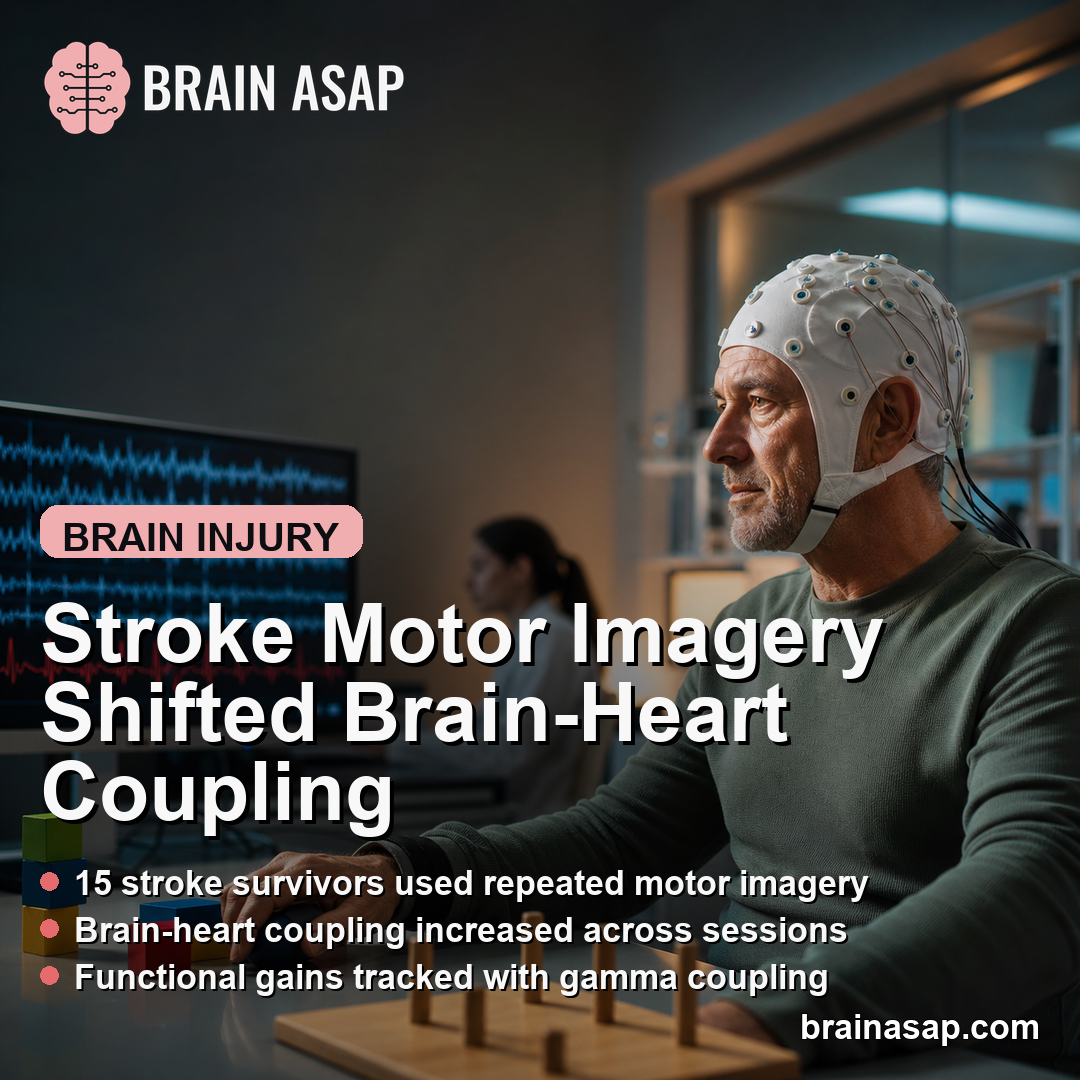
TL;DR: A 2026 preprint in medRxiv found that stroke survivors doing motor imagery showed changing brain-heart coupling across rehabilitation sessions, especially between cardiac sympathetic activity and electroencephalography (EEG) beta-band network clustering.
Key Findings
- 15 stroke survivors: Researchers analyzed two cohorts with upper- or lower-limb motor impairment after stroke.
- 2 motor-imagery protocols: Dataset 1 used gait imagery across 3 sessions; Dataset 2 used hand-grasp imagery across up to 5 visits over 1 year.
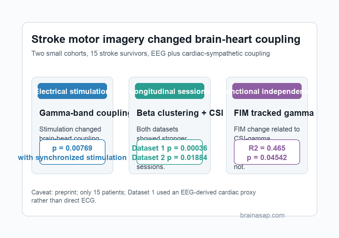
- Electrical stimulation signal: Synchronized stimulation changed cardiac sympathetic index coupling with alpha, beta, and gamma EEG network metrics (p = 0.00769 in several comparisons).
- Beta-band longitudinal effect: Cardiac sympathetic coupling with beta-band clustering increased over sessions in both datasets (p = 0.00036 and p = 0.01884).
- Functional independence link: Functional Independence Measure changes tracked cardiac sympathetic coupling with gamma-band assortativity (R2 = 0.465, p = 0.04542).
Source: medRxiv (2026) | Candia-Rivera et al.
Motor imagery asks a person to imagine a movement without physically performing it. In stroke rehabilitation, the approach can recruit motor planning networks even when the affected limb cannot move enough for ordinary practice.
This preprint focused on a narrower question: whether motor imagery also changes brain-heart interactions, meaning the coupling between EEG network organization and a cardiac sympathetic activity measure derived from heartbeat dynamics.
Stroke Motor Imagery Was Tested Across Two Small Cohorts
The analysis combined 2 independent datasets rather than one large trial. Dataset 1 included 9 stroke patients selected from a larger open dataset because the researchers could recover a usable cardiac proxy from EEG artifact components.
Those patients had hemiplegia confirmed by CT or MRI, were 30 to 70 years old, and had stroke duration from 1 to 12 months. They performed visually guided gait motor imagery across 3 sessions.
- Dataset 1 task: Patients imagined gait phases while viewing visual guidance, with motor imagery alone or motor imagery paired with electrical stimulation.
- Stimulation timing: The affected leg received either invariable electrical stimulation or stimulation synchronized with the gait imagery phase.
- Session spacing: The second session occurred 2 to 4 weeks after the first, and the third occurred 4 to 6 weeks after that.
Dataset 2 included 6 post-stroke patients with hand motor impairment confirmed by MRI. These participants were studied early after stroke, from 5 to 14 days after onset, and returned for longitudinal visits up to 12 months.
The hand-imagery protocol asked patients to imagine closing the affected hand to grasp an object and raise it upward. Researchers also measured neurological status and daily function with the modified Rankin Scale, NIH Stroke Scale, Barthel Index, Functional Independence Measure (FIM), and Fugl-Meyer Assessment.
EEG Networks Were Coupled to a Cardiac Sympathetic Index
The central measurement was not EEG power alone or heart rate alone. Researchers computed time-varying EEG network metrics and compared them with the cardiac sympathetic index (CSI), a heartbeat-variability measure intended to capture slower sympathetic-related rhythm changes.
For the brain side, EEG connectivity was summarized in 3 frequency bands:
- Alpha band: 8.5 to 12 Hz EEG connectivity.
- Beta band: 12.5 to 30 Hz connectivity, a range often relevant to motor-system activity.
- Gamma band: 30.5 to 45 Hz connectivity, used here to track faster network organization during the task.
2 graph measures carried most of the interpretation. Clustering described how strongly network nodes grouped into local neighborhoods, while assortativity described whether nodes with similar connectivity tended to connect with one another.
Brain-heart interaction was quantified with the Maximal Information Coefficient, a method that can capture linear and nonlinear coupling between 2 time series. The reported measure was coordination between systems, not simply higher EEG activity or higher sympathetic activity.
Electrical Stimulation Changed Gamma-Band Brain-Heart Coupling
During the first gait-imagery session in Dataset 1, motor imagery with electrical stimulation produced stronger brain-heart coupling than motor imagery alone. The clearest stimulation result appeared in gamma-band coupling.
With synchronized electrical stimulation, CSI-clustering and CSI-assortativity comparisons were significant across alpha, beta, and gamma bands. Several of those comparisons shared p = 0.00769 after correction.
- Invariant stimulation: The strongest effects involved CSI-assortativity coupling, including gamma-band assortativity.
- Synchronized stimulation: Coupling changed across both clustering and assortativity metrics in alpha, beta, and gamma bands.
- Control checks: Separate brain-only and heart-only metrics did not show the same corrected pattern, which supports a coupling-specific interpretation.
The result fits the idea that sensory stimulation paired with imagined movement can increase task engagement. It does not show that electrical stimulation improved recovery by itself, because the analysis focused on physiological coupling during the task.
Beta-Band Coupling Increased Across Motor-Imagery Sessions
The longitudinal finding was more important for rehabilitation monitoring. Across motor-imagery sessions, CSI coupling with beta-band clustering increased in both datasets.
In Dataset 1, the beta-band CSI-clustering model reported R2 = 0.69, beta = 0.06, and p = 0.00036. In Dataset 2, the corresponding model reported R2 = 0.66, beta = 0.016, and p = 0.01884.
The same beta-band clustering pattern appeared across 2 different protocols.
The gait-imagery dataset and the hand-imagery dataset differed in limb target, timing after stroke, and recording approach, yet the beta-band clustering coupling moved in the same direction.
Other frequency and topology findings were less consistent. Dataset 1 also showed alpha and gamma associations, but Dataset 2 did not reproduce those broadly.
The reproducible longitudinal result was specific: beta-band clustering plus CSI was the most stable signal.
FIM Change Tracked Gamma Assortativity Coupling, Not Every Outcome
Dataset 2 also let researchers compare physiology with clinical change over 1 year. Daily-function measures improved across visits, including the Barthel Index and FIM, while motor impairment measured by the Fugl-Meyer Assessment also improved.
The strongest functional link was narrow. Changes in FIM tracked changes in CSI coupling with gamma-band assortativity, with R2 = 0.465, beta = -0.002, and p = 0.04542.
- FIM association: The significant relationship involved gamma-band assortativity coupled to CSI.
- Barthel result: Barthel changes did not show a corrected significant brain-heart interaction relationship.
- Fugl-Meyer result: Motor-impairment change also did not show a corrected significant relationship with the tested coupling metrics.
- Brain-only and heart-only checks: Separate EEG network or CSI metrics did not explain the clinical-score changes in the same way.
The physiology was not a universal recovery marker. It was a specific coupling measure linked to one daily-function scale in a very small cohort.
Small Sample and ECG Proxy Limit Stroke-Recovery Claims
The main limitation is sample size. The full analysis included only 15 stroke survivors, with varied stroke locations, etiologies, impairment patterns, and missing follow-up sessions.
Dataset 1 also used an EEG-derived cardiac artifact component as a proxy for ECG because direct ECG was not recorded. That proxy made the open dataset usable, but it is not as clean as a direct cardiac recording.
The preprint status also limits clinical use. The work had not been peer reviewed at the time of posting, and the source itself warns that it should not guide clinical practice.
Brain-heart coupling may become a monitoring target for stroke motor-imagery rehabilitation. Larger studies with direct ECG, predefined clinical endpoints, and external validation will need to show whether brain-heart network dynamics can guide brain-computer-interface therapy rather than merely describe recovery physiology.
Citation: DOI: 10.64898/2026.06.23.26355958. Candia-Rivera et al. Motor imagery interventions in stroke patients modulate their brain-heart network dynamics. medRxiv. 2026.
Study Design: Preprint analysis of 2 longitudinal stroke motor-imagery cohorts using EEG network metrics and cardiac sympathetic coupling.
Sample/Model: 15 stroke survivors with upper- or lower-limb motor impairments.
Key Statistic: CSI coupling with beta-band clustering increased across sessions in both datasets, with p = 0.00036 in Dataset 1 and p = 0.01884 in Dataset 2.
Caveat: Small preprint study with heterogeneous stroke patients, missing sessions, and an EEG-derived cardiac proxy in one dataset.