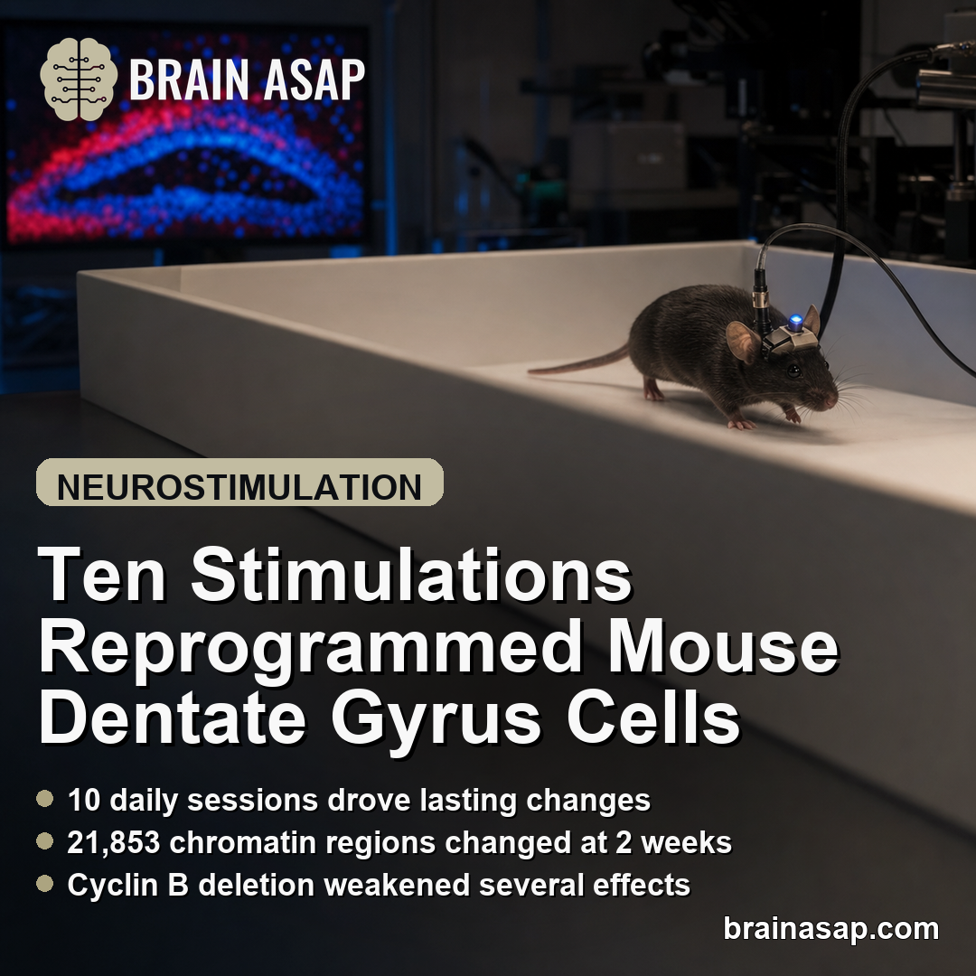
TL;DR: A 2026 mouse study in Nature Communications reported that 10 daily bouts of optogenetic dentate-gyrus stimulation produced weeks-long changes in chromatin, nuclear structure, behavior, and spatial coding, while Cyclin B deletion weakened several effects; this ECT-inspired model does not establish how electroconvulsive therapy works in people.
Key Findings
- Ten stimulation sessions produced the durable molecular response: Mice received five minutes of dentate-gyrus stimulation per day. Changes after three days largely receded, while many changes after 10 days remained two weeks to more than one month later.
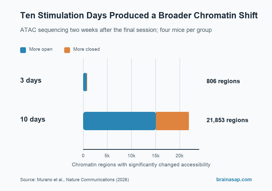
- Chromatin accessibility changed at 21,853 regions: Two weeks after the 10-day protocol, ATAC sequencing detected 15,098 more-open and 6,755 more-closed regions. The corresponding three-day group had 806 altered regions.
- Behavioral changes were mixed: The 10-day group showed less immobility and shorter feeding latency in stress-based tests, but also greater locomotion, impaired remote memory, reduced spatial coding, and no significant change in sucrose preference.
- Cyclin B deletion weakened several lasting effects: Removing Cyclin B1 and B2 from dentate-gyrus cells reduced nuclear, cell-maturity, and prolonged locomotor changes, although it did not fully prevent every response.
- The human comparison was small and observational: Postmortem gene-expression data included seven patients with an ECT history and 61 without one. Similarity to the mouse expression pattern does not prove a shared treatment mechanism.
Source: Murano et al., Nature Communications (2026).
Electroconvulsive therapy (ECT) and repetitive transcranial magnetic stimulation (rTMS) expose the brain to very different forms of stimulation, yet both can produce effects that outlast a treatment session. The cellular events that maintain those effects remain uncertain.
Researchers built a precise mouse model called REPetitive OPtogenetic Stimulation (REPOPS), using light-sensitive channels to activate granule cells in the hippocampal dentate gyrus. This is a targeted laboratory model of repeated neural activation, not a miniature version of clinical ECT.
Ten Daily Stimulations Produced Weeks-Long Molecular Changes
Adult mice received 10-millisecond light pulses at 10 hertz for five minutes per day. Researchers compared three consecutive stimulation days with 10 days, then collected tissue 24 hours or two weeks after the final session.
Calbindin, a marker of mature dentate-gyrus granule cells, decreased after both schedules. It returned toward control levels within two weeks after three sessions but remained lower after 10 sessions, indicating a more persistent shift in cellular state.
RNA sequencing used six mice per group. Gene-expression changes after the three-day protocol diminished over time, whereas the 10-day response remained broad at two weeks and resembled an immature dentate-gyrus expression pattern.
ATAC sequencing, which measures how accessible DNA regions are to regulatory machinery, used four mice per group. At two weeks, the three-day schedule was associated with 806 altered chromatin regions: 748 more open and 58 more closed.
The 10-day schedule was associated with 21,853 altered regions: 15,098 more open and 6,755 more closed. The count shows a much broader molecular response, not 21,853 proven causes of a behavioral change.
Dentate-Gyrus Neurons Entered a Partial G2/M-Like State
The persistent expression pattern included genes normally associated with the G2/M portion of the cell cycle, including Cyclin B1, Cyclin B2, Cyclin A2, and Cdc25c. Cyclin B helps cells move from the G2 growth phase toward mitosis.
Mature neurons generally do not divide. Here, stimulation was followed by Cyclin B expression, disruption of the lamin B1 nuclear boundary, more phosphorylated histone H3, and larger chromocenters, which are condensed regions of DNA inside the nucleus.
These measurements came from three mice per group in several histology analyses, with hundreds of cells evaluated. The statistical models treated mouse identity as a random effect so that many cells from one animal were not counted as fully independent animals.
The evidence supports a partial G2/M-like program, not actual neuronal division. Researchers found no fully compacted chromosomes, the nuclear changes were milder than true mitosis, and the stimulation protocol did not produce significant cell death.
Locomotion, Stress Tests, Memory, and Spatial Coding All Changed
Ten-day stimulation increased open-field activity for at least two weeks and home-cage activity for more than one month. Main behavioral experiments generally used six to 10 mice per group, with seven to 16 per group across the full behavioral program.
Two weeks later, 10-day mice spent less time immobile in the tail-suspension test and began eating sooner in the novelty-suppressed feeding test than unstimulated or three-day mice. An analysis adjusting feeding latency for locomotor activity still found a stimulation-group difference.
Greater movement complicates interpretation of immobility-based tests. The forced-swim result was weaker, sucrose preference did not differ significantly, and the protocol did not produce a uniform antidepressant-like profile.
Potential costs included impaired remote memory in a supplemental experiment and a shift in dentate-gyrus coding away from position and toward speed. Calcium-imaging analyses used five mice per group; position decoding worsened while speed decoding improved for more than two weeks.
Those mixed outcomes matter for the ECT analogy. Reduced immobility cannot be separated from increased movement by rhetoric alone, and a mouse stress test is not a diagnosis or a measure of depression recovery in a patient.
Cyclin B Deletion Reduced Nuclear and Lasting Behavioral Effects
Researchers used viral CRISPR tools to remove Cyclin B1 and Cyclin B2 from dentate-gyrus cells before the 10-day stimulation schedule. A scrambled guide sequence served as the control.
Cyclin B deletion reduced the stimulation-related changes in lamin B1, phosphorylated histone H3, chromocenter size, and calbindin. It also prevented the prolonged locomotor increase from remaining significant on day 24 in an experiment with seven mice per group.
The knockout did not erase every effect: lamin B1 and calbindin still changed after stimulation, but less than in control-edited mice. That pattern places Cyclin B within the mechanism without making it the only pathway involved.
Cyclin B was also needed to maintain higher levels of ΔFosB, a long-lived activity-regulated transcription factor, at two weeks. Pharmacologic manipulation of Cyclin B/Cdk1 signaling moved ΔFosB and calbindin in complementary directions, adding mechanistic support within this mouse system.
Seven ECT-Exposed Postmortem Samples Cannot Validate a Human Mechanism
Researchers reanalyzed postmortem dentate-gyrus RNA sequencing from people with major depressive disorder or bipolar disorder. The dataset included seven patients with an ECT history, 61 patients without that history, and 93 healthy controls.
Genes that differed by ECT history overlapped with developmental gene-expression changes and with several synapse-related changes in stimulated mice. This provides a human point of comparison, but exposure was not randomized and the dataset cannot separate ECT from diagnosis, illness severity, other treatments, or postmortem factors.
The human analysis measured gene expression only. It did not test Cyclin B deletion, nuclear structure, neural coding, memory, or treatment response, and seven exposed samples provide limited precision.
Mouse omics experiments used males only, while some behavioral and histology experiments included both sexes. Optogenetic activation of one hippocampal cell population also differs sharply from ECT or rTMS in stimulation pattern, brain coverage, and clinical context.
The results identify a testable sequence in mice: repeated calcium entry, partial cell-cycle-like nuclear remodeling, persistent ΔFosB, and altered cellular and circuit function. Human studies would need to measure those steps directly before this pathway could be treated as an ECT mechanism or therapeutic target.
Citation: DOI: 10.1038/s41467-026-74202-w. Murano et al. Repetitive neuronal activation regulates cellular maturation state via nuclear reprogramming. Nature Communications. 2026;17:5881.
Study Design: Repeated optogenetic stimulation experiments in mice, with molecular, histologic, behavioral, and calcium-imaging assays plus secondary analysis of human postmortem RNA sequencing.
Sample/Model: ATAC sequencing used four mice per group, calcium imaging used five, and main behavioral figures used six to 10; the Methods reports seven to 16 per group across the broader behavioral set.
Key Statistic: Two weeks after stimulation, ATAC sequencing detected 21,853 altered chromatin regions after the 10-day protocol versus 806 after the three-day protocol.
Caveat: Targeted optogenetic stimulation and mouse behavioral assays cannot establish the mechanism, effectiveness, or safety of clinical ECT in people.