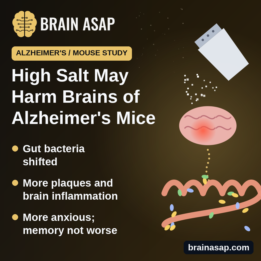
TL;DR: Alzheimer’s-model mice fed very salty chow for 6 months had altered gut bacteria, more brain inflammation and plaques, and more anxious behavior than mice on normal chow. It’s a mouse study, and the gut-to-brain link was not proven.
Key Findings
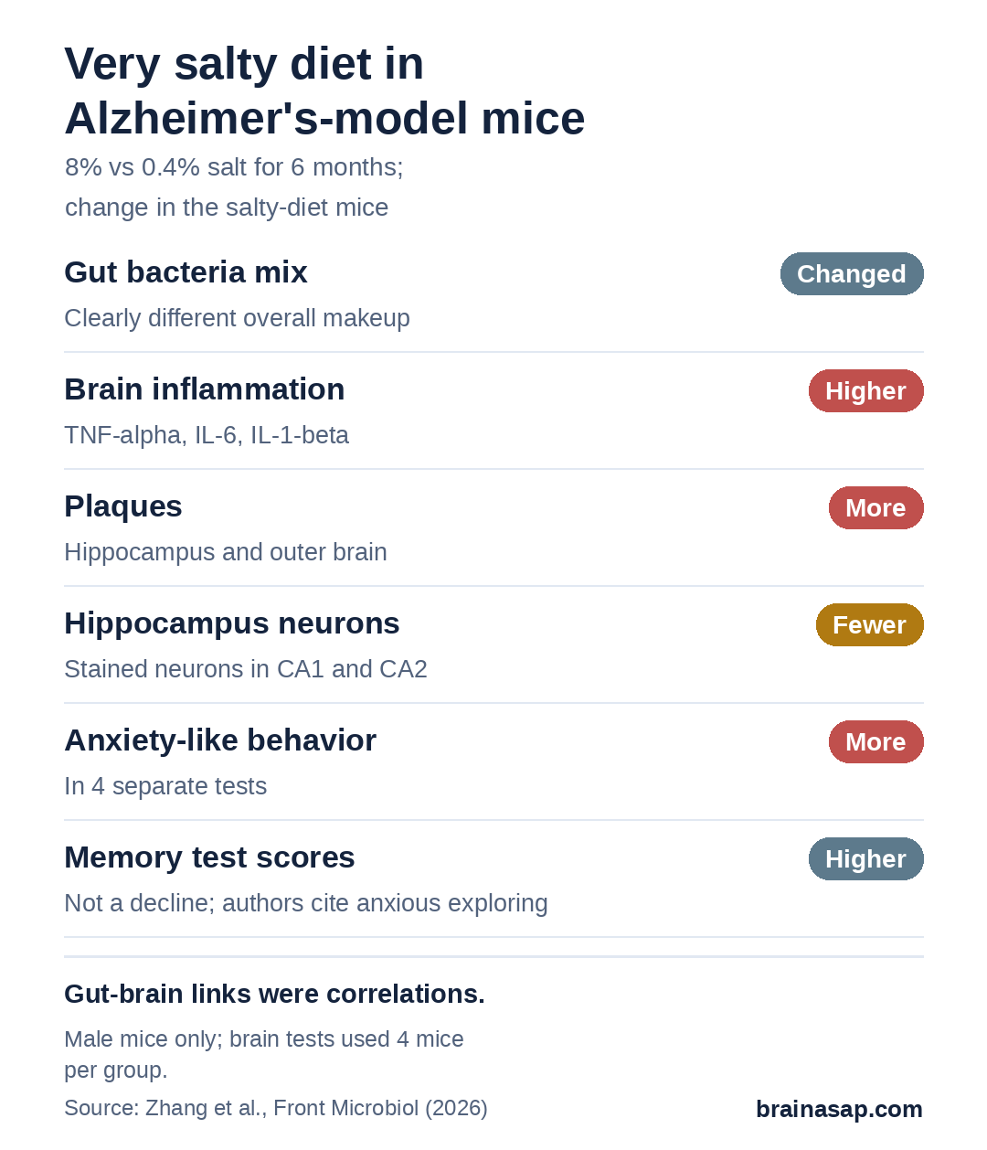
- 8% vs 0.4% salt for 6 months: Male Alzheimer’s-model mice, about 40 per diet group overall.
- Gut bacteria shifted: Overall mix of gut microbes differed clearly from mice on normal chow.
- Brain changes: More plaques, more inflammation markers and fewer intact neurons in the hippocampus.
- More anxious behavior: Less time in open, bright spaces and more marble burying.
- Memory tests did not show decline: Scores actually went up, which the authors put down to anxious exploring.
Source: Frontiers in Microbiology (2026) | Zhang et al.
Most people eat more salt than health guidelines recommend, and too much salt is already linked to high blood pressure and stroke. Researchers are now asking whether it also affects the brain, possibly through the bacteria living in the gut.
A team from several hospitals and universities in China fed a very salty diet to mice that develop Alzheimer’s-like disease, then looked at their guts, brains and behavior.
Six Months on Very Salty Chow
The study used male APP/PS1 mice, a common lab strain engineered to build up amyloid plaques like those seen in Alzheimer’s disease. Starting at 6 months old, mice were randomly assigned to one of two diets:
- Normal chow: 0.4% salt, 43 mice.
- High-salt chow: 8% salt, 20 times more, 41 mice.
Not every mouse went through every test. Behavior tests used 10 mice per group, and brain tissue tests used 4 per group.
Salty-diet mice drank more water, weighed less by months 5 and 6, and had higher diastolic (lower-number) blood pressure.
Plaques, Inflammation and Neuron Loss in the Brain
Compared with normal-chow mice, salty-diet mice showed:
- More plaques in the hippocampus and the outer brain.
- Fewer stained neurons in two parts of the hippocampus (CA1 and CA2), the brain’s memory hub.
- Fewer branches on the neurons’ receiving ends.
- Higher inflammation signals, including TNF-alpha, IL-6 and IL-1-beta, in the brain and liver.
Gut Bacteria Shifted, and Some Tracked With Brain Chemistry
The overall makeup of the gut community differed clearly between the diets, with a higher ratio of two major bacterial groups, Firmicutes to Bacteroidota. Several gut bacteria families rose or fell in step with chemicals in the hippocampus, such as glutamate, a key messenger for learning.
Those are correlations. The study did not test whether changing the gut bacteria changes the brain, and it did not measure whether the gut lining became leaky.
More Anxious, but Memory Tests Went the Other Way
Salty-diet mice behaved in more anxious ways across four tests: they avoided the open center of a box, stayed in enclosed arms of a raised maze, buried more marbles and ventured less into a bright compartment.
On two memory-related tests, though, salty-diet mice scored higher, not lower: they explored a new object more and alternated between maze arms more often. The authors argue this reflects anxiety-driven exploring rather than better memory. Either way, these tests do not show memory decline, even though the paper’s summary describes worsened cognition.
What Mouse Salt Doesn’t Tell Us About People
- Extreme dose: 8% salt chow has no direct match to a human diet.
- Males only, in one engineered strain.
- Tiny tissue groups: 4 mice per group for brain and inflammation tests.
- Link, not mechanism: Gut changes were associated with brain changes, not shown to cause them.
Would Restoring Gut Bacteria Protect the Brain?
The study adds to animal evidence that a very salty diet can affect the brain in an Alzheimer’s model, with gut bacteria as one possible route. The next step is to test that route directly, for example by transferring gut bacteria between mice or reversing the shift, and to see whether more realistic salt levels matter.
Citation: DOI: 10.3389/fmicb.2026.1884427. Zhang H, Kou J, Ge P, Gong L, Zhang W, Fei Y. Gut microbiota dysbiosis is associated with hippocampal neuronal damage and behavioral alterations in APP/PS1 mice with high-salt diet. Front Microbiol. 2026;17:1884427.
Study Design: Randomized diet experiment in male APP/PS1 transgenic mice, with gut sequencing, brain histology, inflammation assays, metabolomics and behavior tests.
Sample Size: 84 mice (43 normal diet, 41 high-salt); assay subsets of 4 to 13 per group.
Key Statistic: Reduced Nissl-stained neurons in hippocampal CA1 (p < 0.001) and CA2 (p < 0.05) with high salt.
Caveat: Mouse study; extreme salt dose; gut-brain links are correlational; memory tests did not show decline.