Brainstem Expiratory Neurons Drove Hypertension
TL;DR: A brainstem region involved in forceful exhalation also appears to drive blood-vessel constriction, making it a possible target for some forms of neurogenic hypertension.
Key Findings
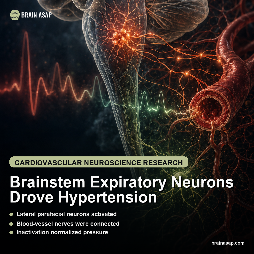
A forced-exhalation region joined the pressure mechanism: The lateral parafacial region, normally recruited for strong expiration, was active under hypertensive conditions.
Blood-vessel nerves were part of the route: The region connected to sympathetic pathways that tighten blood vessels and raise pressure.
Silencing the region lowered pressure: Inactivating the lateral parafacial region brought blood pressure back to normal levels in the reported model.
The carotid bodies offered a peripheral handle: Oxygen-sensing clusters in the neck activated the brainstem region, suggesting a possible way to influence it without broadly drugging the brain.
Sleep apnea is the key patient context: Because carotid bodies activate when breathing stops at night, the pathway matters most for testing in people with sleep-disordered breathing and hypertension.
Source: Circulation Research (2026) | Magalhaes et al.
High blood pressure is usually discussed as a problem of arteries, kidneys, salt, hormones, or stress. This paper points somewhere less obvious: a brainstem breathing circuit that can help keep blood vessels tightened in some forms of hypertension.
The Lateral Parafacial Region Linked Forced Exhalation to Blood Pressure
The lateral parafacial region sits in the brainstem, among the circuits that handle automatic jobs such as breathing, heart rate, and digestion. This is not the breath-control system people use when they decide to slow down and relax. It is deeper, older physiology.
The lateral parafacial region is recruited during forced exhalation: coughing, laughing, exercise, or any hard breath out that pulls in the abdominal muscles. Quiet exhalation is different. Most of the time, the lungs simply recoil.
The surprise is that the same region also connects with nerves that tighten blood vessels. In other words, a circuit that helps push air out of the lungs may also help push blood pressure up.
That link is easy to miss because breathing and blood pressure are often discussed in separate rooms: pulmonology over here, cardiology over there, neuroscience somewhere else entirely. The brainstem does not respect those boundaries. It coordinates oxygen, circulation, and muscle activity in the same cramped biological neighborhood.
Blood Vessel Tightening Is How the Brainstem Can Raise Pressure
Blood pressure rises when blood has to move through a tighter, more resistant vascular system. Sympathetic nerves can create that state quickly by telling blood vessels to constrict.
The body needs fast vascular control during exercise, threat, posture changes, and oxygen stress. A reflex that helps in those moments can cause trouble if it stays switched on or gets recruited by disease.
That is the cardiovascular relevance of the lateral parafacial region. The finding connects a breathing-related brainstem signal to the same sympathetic output that can keep blood vessels narrowed.
The paper argues that active-expiration neurons feed into sympathetic vasoconstriction. Vasoconstriction means blood vessels narrow, which raises resistance and can push blood pressure higher. If that pathway is overactive, hypertension can be partly driven from the brainstem rather than only from the vessels themselves.
2 patients can have the same cuff reading for very different reasons: kidney signaling in one, vascular stiffness in another, excessive sympathetic drive in a third. This study gives researchers a candidate circuit for the sympathetic-drive version.
Inactivating the Lateral Parafacial Region Lowered Pressure
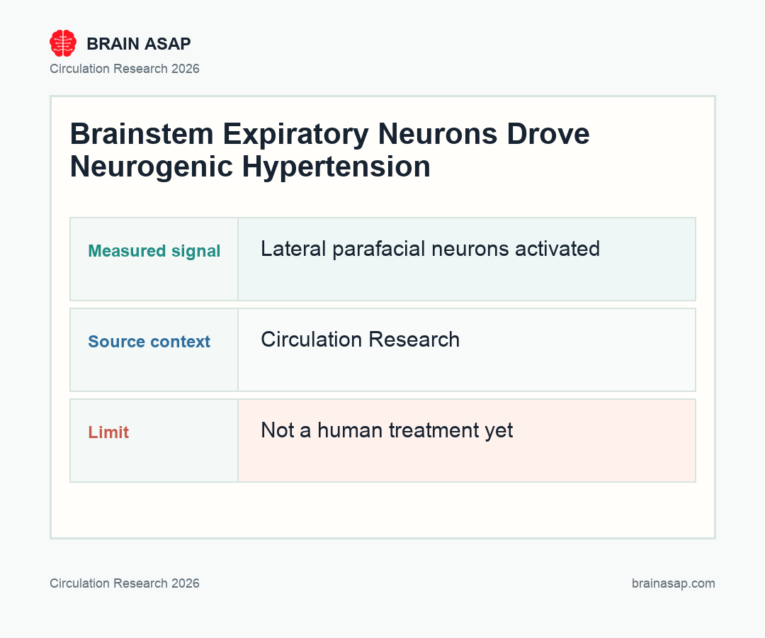
The strongest result is the inactivation finding. Under hypertensive conditions, the lateral parafacial region was activated. When the researchers inactivated the region, blood pressure fell to normal levels.
This circuit is not an all-purpose explanation for high blood pressure. Hypertension has many causes. But the inactivation result moves the finding beyond a loose association: the region was active and functionally involved; in the reported model, it helped sustain the hypertensive state.
The pathway is worth testing in more detail. A brainstem signal tied to both forced breathing and vessel constriction is specific enough to manipulate, compare, and eventually look for in more defined patient groups.
Carotid Bodies Could Offer a Route Around Direct Brainstem Drugs
A small brainstem region is a difficult therapeutic target. A drug that enters the brain can affect many regions at once, and the brainstem is not a place where broad side effects are acceptable.
The more realistic opening came from the carotid bodies, small oxygen-sensing clusters in the neck near the carotid arteries. These sensors can activate the lateral parafacial region from outside the brain.
Instead of trying to drug the brainstem directly, researchers can test whether quieting carotid-body activity reduces the drive into the lateral parafacial region.
For now, that is a research path, not a treatment recommendation. But it is a cleaner therapeutic idea than “the brain is involved”: target the neck-based oxygen sensors that feed the pressure-raising brainstem circuit.
The caution is just as important. Carotid bodies help the body detect oxygen and respond to respiratory stress, so any future intervention would need to reduce harmful overactivity without impairing a protective oxygen-sensing response.
Sleep Apnea Could Recruit the Same Oxygen-Sensing Pathway
The sleep-apnea connection follows directly from the carotid bodies. When breathing stops during sleep, oxygen-sensing pathways can fire repeatedly. Many people with sleep apnea also show elevated sympathetic tone and high blood pressure.
If carotid-body activation can recruit the lateral parafacial region, the pathway is especially relevant in people whose hypertension travels with disordered breathing at night. That is not proof of a human treatment target, but it is a plausible place to look.
The clinical idea is narrow. Some patients with high blood pressure, abdominal breathing patterns, or sleep-disordered breathing can have a neural driver that routine blood-pressure categories do not capture.
This does not replace standard blood-pressure care. It can help explain why the same diagnosis hides several biological routes.
It also gives clinicians and researchers a sharper question to ask. Instead of treating sleep apnea as a separate comorbidity sitting beside hypertension, future studies can ask whether repeated oxygen-sensor activation is helping push the pressure itself.
The Finding Points to a Blood-Pressure Target, Not a Treatment Yet
This finding should not be read as advice to stop standard hypertension care, change medication, or treat breathing exercises as a substitute for medical management. Blood pressure is high-stakes physiology, and most patients need ordinary evidence-based risk control.
The paper adds a breathing-linked brainstem region to the machinery that can raise vascular tone, at least in the studied model. That can help explain why some hypertension looks neurogenic, why sleep apnea and blood pressure often travel together, and why a one-size-fits-all approach can miss the driver.
The next questions are concrete:
- Which patients show this pathway? The relevant group is likely defined by sleep apnea, abdominal breathing, carotid-body overactivity, neurogenic hypertension, or some combination.
- Can carotid-body targeting safely quiet it? The appeal is peripheral access, but oxygen sensing is important physiology and would need careful control.
- How selective is the circuit? A brainstem node that participates in breathing and pressure control must be manipulated without disrupting essential automatic functions.
- What are the exact model details? The accessible source does not provide full animal counts, blood-pressure values, or intervention parameters, so the full paper remains necessary before publication-level precision.
The result shows that a forced-expiration circuit is now a candidate target for blood-pressure control in patients whose hypertension is being pushed by sympathetic drive.
Paper: Lateral Parafacial Neurons Evoked Expiratory Oscillations Driving Neurogenic Hypertension. Circulation Research. 2026. DOI: 10.1161/CIRCRESAHA.125.326674
Authors: Magalhaes et al.
Study Design: Mechanistic cardiovascular-neuroscience study of lateral parafacial brainstem neurons, carotid-body input, expiratory oscillations, sympathetic output, and blood pressure control.
Sample Size: Experimental hypertension models; exact animal/sample counts require full-text confirmation.
Key Statistic: The accessible source reports that inactivating the lateral parafacial region lowered blood pressure to normal levels in the hypertensive model.
Important Caveat: The accessible source does not provide all animal counts or intervention details, so publication-level precision requires checking the full paper.