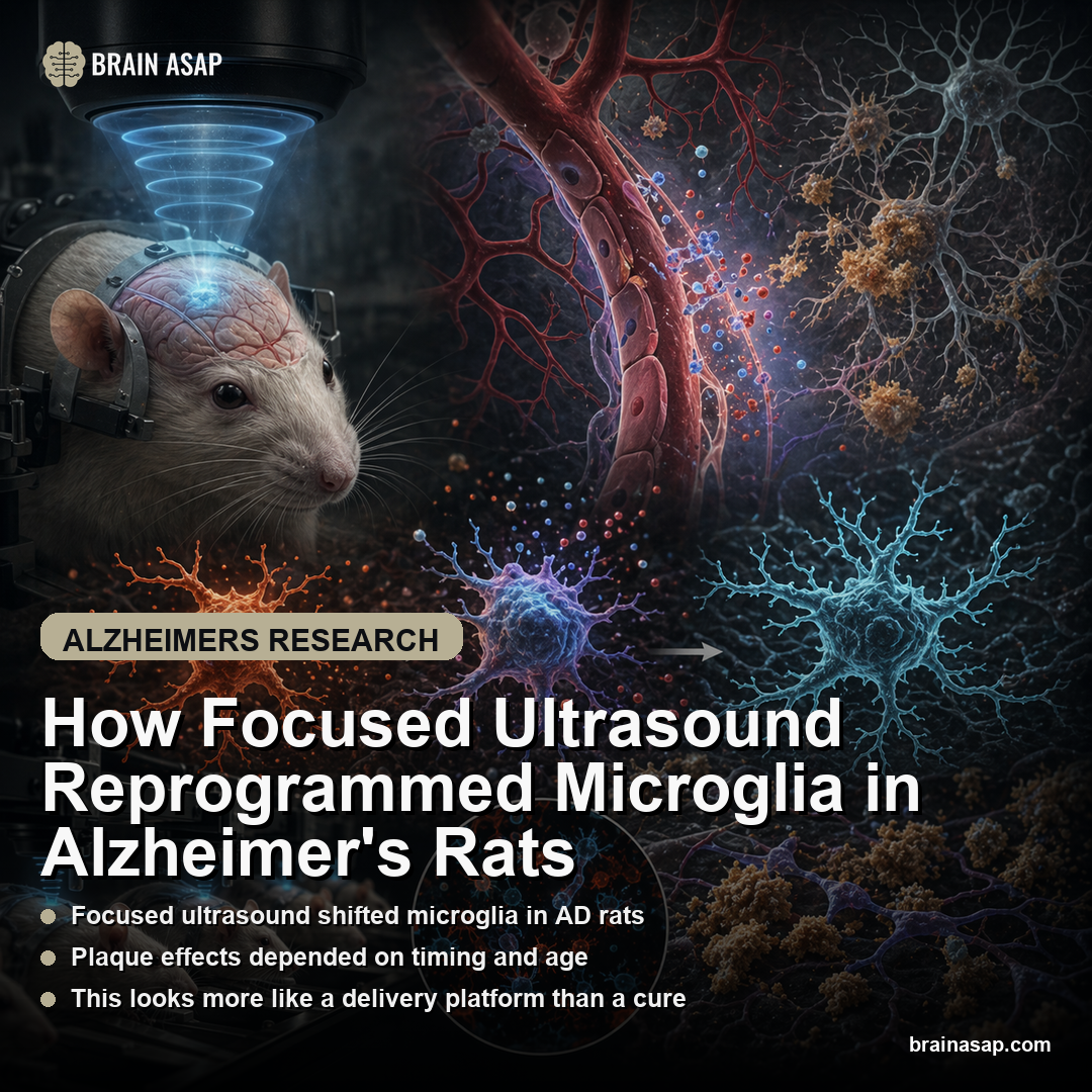
How Focused Ultrasound Reprogrammed Microglia in Alzheimer’s Rats
TL;DR: In TgF344-AD rats, repeated low-intensity focused ultrasound triggered a short-lived inflammatory response, then left behind a more durable microglial metabolic shift in early disease without clearly improving Alzheimer’s pathology on its own.
Key Findings
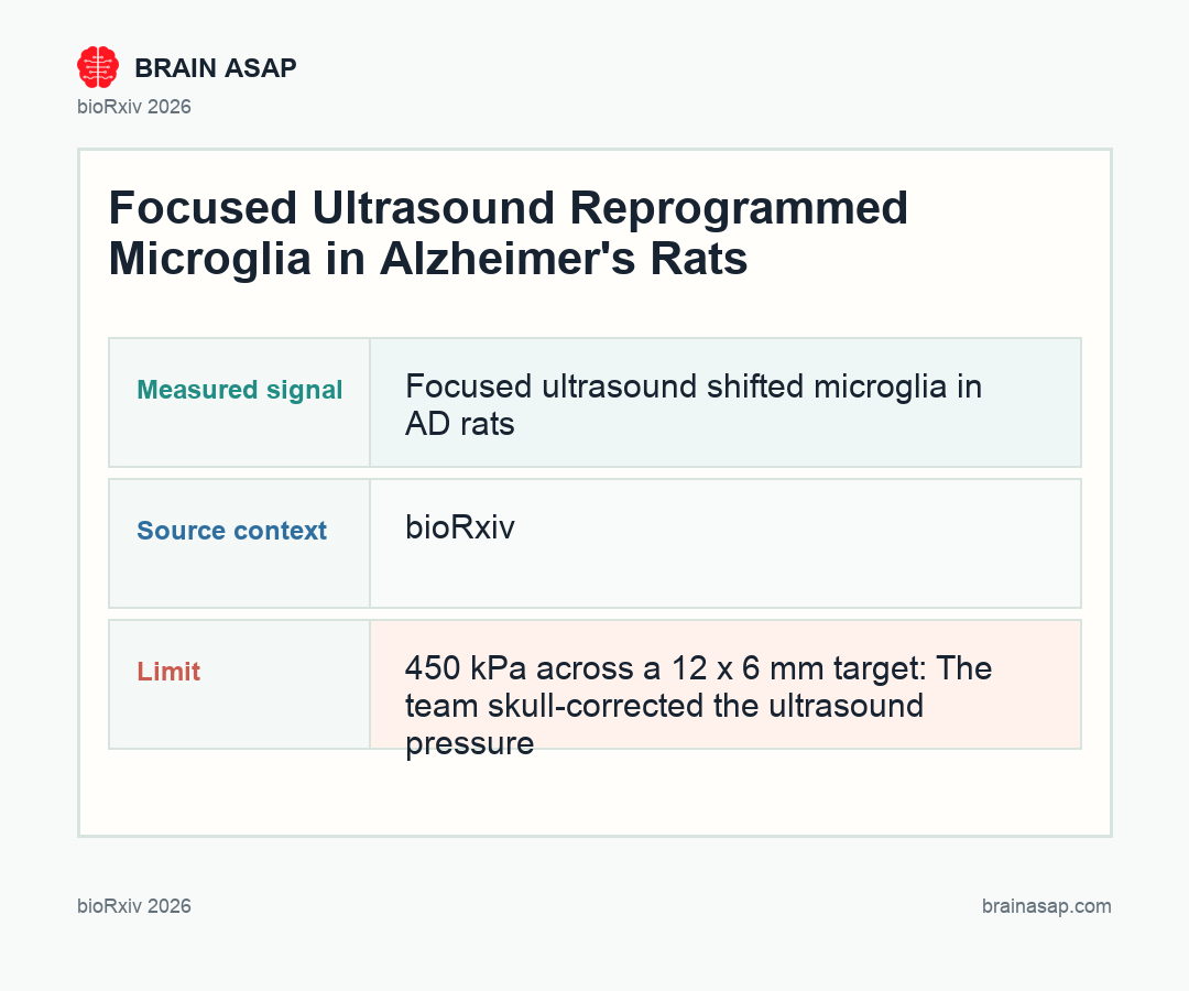
- 450 kPa across a 12 x 6 mm target: The team skull-corrected the ultrasound pressure and swept a large bilateral brain volume in TgF344-AD rats, making this a fairly aggressive blood-brain barrier opening protocol rather than a tiny focal proof of concept.
- Day-1 hemorrhages cleared by day 7: Small post-treatment microhemorrhages were visible 24 hours after sonication, but they were no longer present a week later, and four weekly sessions did not appear to stack that injury signal over time.
- Single exposure lowered aggregated Aβ42: One LiFUS plus microbubble session in older rats reduced the highly aggregated guanidine-soluble Aβ42 fraction within 24 hours (p = 0.0226) without changing pTau231.
- Four weekly sessions briefly worsened pathology markers: 7 days after the fourth treatment in older rats, highly aggregated Aβ42 rose instead of fell (p = 0.0059) and pTau231 was also higher (p = 0.0009), alongside a microglia-heavy inflammatory transcriptional response.
- Old rats mostly normalized by 6 weeks: At the later follow-up, repeated treatment no longer changed Aβ42 or pTau231, while guanidine-soluble Aβ40 was lower (p = 0.0138) and most sorted-microglia readouts were no longer meaningfully altered.
- Young-stage rats showed microglial rewiring: When the same four-session protocol was started earlier, TSPO fell in total parenchyma (p = 0.0314) and sorted microglia (p < 0.0001), while Tnfα rose and Glut1 increased, consistent with a shifted inflammatory and metabolic state rather than obvious plaque clearance.
Source: bioRxiv (2026) | Abjean et al.
Focused ultrasound is often pitched as a way to pry open the blood-brain barrier and help Alzheimer’s drugs reach the tissue they are supposed to treat. This preprint asks a harder question: what happens if you repeatedly open that barrier in an Alzheimer’s brain even before you add a drug?
Why Alzheimer’s Drug Delivery Keeps Colliding With the Blood-Brain Barrier
Alzheimer’s drug development has a geometry problem. Antibodies such as lecanemab and donanemab can bind amyloid, but the blood-brain barrier makes it hard to get enough drug into the brain without giving large systemic doses that raise the risk of side effects.
That is why low-intensity focused ultrasound has attracted so much attention. Pair it with intravenous microbubbles, and the oscillating bubbles temporarily loosen the barrier. In theory, that lets researchers deliver more treatment to the brain with less drug in the bloodstream.
But barrier opening is not biologically neutral. It physically perturbs vessels, glia, and the inflammatory environment. The study is important because it asked what that perturbation looks like in an Alzheimer’s model even before the technology is paired with a therapeutic payload.
What One 24-Hour Ultrasound Session Did to Aggregated Aβ42
The first surprise came from the single-treatment arm in 18- to 19-month-old TgF344-AD rats. Within 24 hours, the procedure slightly nudged inflammatory genes upward, including Aif1, Gfap, Vimentin, Tspo, Il6, and Rantes, but the tissue-level protein stains for IBA1 and glial fibrillary acidic protein (GFAP), an astrocyte protein that can rise with brain injury or inflammation did not move enough to show an obvious morphologic response.
Pathology readouts were mixed rather than broadly beneficial. The highly aggregated guanidine-soluble Aβ42 fraction fell significantly after one exposure, but Aβ40 did not budge and pTau231 stayed flat. That suggests the earliest effect was selective, fast, and probably tied to how different amyloid pools respond to barrier opening rather than a general reset of Alzheimer’s pathology.
The safety profile was also nuanced. Microhemorrhages were visible at 24 hours, which is not trivial in an amyloid-laden brain, but they had resolved by day 7 and did not obviously worsen with repeated weekly treatment. That is reassuring for the technology, though it still means the procedure creates a real acute vascular insult that cannot be waved away as harmless.
Four Weekly Sessions Looked Worse at Day 7 Than at Week 6
The most counterintuitive result in the paper is that more ultrasound was not simply better ultrasound. In older rats, four weekly LiFUS plus microbubble sessions produced a persistent inflammatory transcriptional signature 7 days after the last treatment, and that signal leaned heavily toward microglia.
This was the time point where the pathology markers looked least encouraging. Aggregated Aβ42 increased instead of decreased, and pTau231 also rose. Those are exactly the wrong directions if you were hoping repeated barrier opening alone would steadily chip away at Alzheimer’s hallmarks.
The pattern changed again 6 weeks later. By then, the short-term inflammatory signature had largely faded, Aβ42 and pTau231 were no longer different, and only guanidine-soluble Aβ40 remained lower. The result is less a clean therapeutic arc than a rebound pattern: a short-term disturbance, followed by partial normalization, with no convincing sign that ultrasound by itself meaningfully changed the disease trajectory in aged animals.
- At 24 hours: one session lowered aggregated Aβ42 but produced a small acute inflammatory signal.
- At day 7 after repeated sessions: older rats showed the least favorable pathology pattern.
- By week 6: much of the disturbance had faded without a clear standalone disease-modifying effect.
How Younger Alzheimer’s Rats Showed Microglial Rewiring Instead of Plaque Clearance
The early-stage cohort is the most important part of the study. When the researchers started the same four-session protocol in younger, pre-symptomatic rats, the treatment still did not clearly reduce amyloid or tau. But it did change the biology of the microglia in a way that looked more like reprogramming than acute activation.
TSPO dropped in both total parenchyma and sorted microglia, P2y12r fell, Tnfα rose, and Tgfβ trended upward. That is not a neat pro-inflammatory or anti-inflammatory interpretation. The authors explicitly push against that binary framing and instead argue for a continuum model in which microglia adapt their state to the changed vascular and metabolic environment.
There were metabolic hints too. Glut1 increased, Mct1 trended down, and Vdac1 also trended lower, a pattern the authors interpret as a shift toward glycolytic metabolism. In plain language: repeated barrier opening may be nudging microglia away from one energy strategy and toward another.
Lower TSPO and Higher Glut1 Hint at a Metabolic Microglial Shift
That metabolic angle is what keeps this paper from collapsing into a simple “ultrasound caused inflammation” story. TSPO is often treated as a marker of neuroinflammation, but here it moved in opposite directions depending on timing. It rose after acute exposure, then fell weeks later after repeated treatment, especially in younger animals.
The authors argue that this split makes biological sense. The early TSPO rise looks like a transient inflammatory response to barrier opening. The later TSPO reduction, paired with shifts in Tnfα, Glut1, and mitochondrial genes, looks more like a sustained functional adaptation in microglia.
That is an important distinction for Alzheimer’s research. If focused ultrasound is eventually used as a drug-delivery platform, its value may depend not just on how much of the barrier it opens, but on whether repeated exposures are quietly reshaping the immune-metabolic state of the brain around the therapy.
This Still Looks More Like a Delivery Platform Than a Standalone Alzheimer’s Therapy
The paper’s final message is more restrained than the field’s hype. In these rats, repeated LiFUS plus microbubbles did not reliably improve the core Alzheimer’s pathology markers on its own. It produced some potentially useful biological effects, but they were conditional on disease stage and time point, and several of the short-term changes actually moved in the wrong direction.
That does not make the approach a failure. It may simply clarify where the technology is strongest.
Focused ultrasound still looks promising as a drug-delivery tool that could reduce the systemic dose needed for antibodies or other therapeutics. What this study does not support is the stronger claim that repeated barrier opening by itself is already an Alzheimer’s treatment.
That is the judgment readers should carry away. The interesting signal here is not plaque removal. It is that microglia may be one of the main long-term responders to repeated ultrasound-mediated barrier opening, especially early in disease, and that any future therapeutic strategy will probably have to account for that biology rather than treating the ultrasound step as passive plumbing.
Paper: Repeated low-intensity focused ultrasound induces microglial profile changes in the TgF344-AD rat model of Alzheimer's disease. bioRxiv. 2026.. DOI: 10.1101/2024.09.25.614692
Authors: Abjean et al.
Study Design: Cohort study
Sample Size: 450 kPa across a 12 x 6 mm target: The team skull-corrected the ultrasound pressure and swept a large bilateral brain volume in TgF344-AD rats, making this a fairly aggressive blood-brain barrier opening protocol rather than a tiny focal proof of concept.
Key Statistic: Day-1 hemorrhages cleared by day 7: Small post-treatment microhemorrhages were visible 24 hours after sonication, but they were no longer present a week later, and four weekly sessions did not appear to stack that injury signal over time.