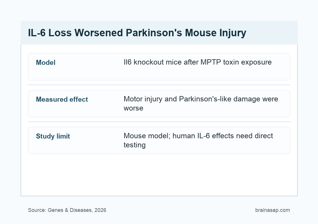
TL;DR: A 2026 study in Genes & Diseases found that Il6 deficiency, meaning loss of the gene for interleukin-6, worsened Parkinson’s-like motor and dopamine-system injury in mice.
Key Findings
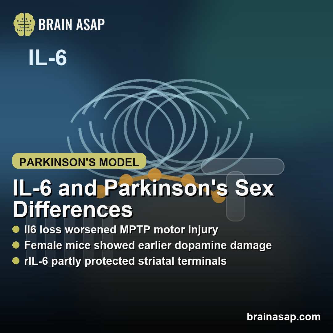
- Il6 knockout worsened movement: Mice lacking Il6 performed worse after MPTP, a toxin commonly used to model Parkinson’s-like dopamine injury.
- Female knockout mice showed earlier damage: Dopamine fiber loss, substantia nigra neuron loss, and astrocyte activation were more pronounced at the early 3-day time point.
- An alpha-synuclein model pointed the same way: Female Il6 knockout mice developed earlier motor dysfunction and worse dopamine neurodegeneration after A53T alpha-synuclein overexpression.
- Male injury appeared later: Male knockout mice showed clearer dopamine-neuron differences at the later time point, making the IL-6 effect sex- and time-dependent rather than uniform.
- Recombinant IL-6 partly rescued the model: 500 ng rIL-6 injections partially improved motor behavior and striatal dopamine-terminal preservation in MPTP-treated mice.
Source: Chen et al. Genes & Diseases. 2026.
Interleukin-6 is usually discussed as an inflammatory signal, but this mouse study argues that its role in Parkinson’s biology is not simply harmful.
Researchers tested what happened when the Il6 gene was removed, then challenged mice with 2 different Parkinson’s-like injury models.
The main result was direct: Il6 deficiency made the Parkinson’s-like phenotype worse. The effect appeared in both sexes, but the timing and tissue pattern were not identical.
Female knockout mice showed earlier and broader injury after MPTP, while male knockout mice showed a clearer dopamine-neuron difference later.
The sex difference is clinically relevant because Parkinson’s disease is sexually dimorphic in humans. Men have higher disease prevalence, while women can show different symptom patterns, hormone interactions, and progression features.
A mouse model cannot settle the human question, but it can test whether an inflammatory pathway behaves differently by sex under controlled injury conditions.
Researchers Tested IL-6 Loss in Two Parkinson’s Mouse Models
The study used Il6 knockout mice, Il6 heterozygous mice, and wild-type mice on a C57BL/6 background. Knockout mice lacked both copies of the gene; heterozygous mice retained 1 copy.
Researchers then used 2 Parkinson’s-like models:
- MPTP toxin model: mice received 15 mg/kg MPTP 4 times at 2-hour intervals to injure the nigrostriatal dopamine system.
- Alpha-synuclein A53T model: female mice received an AAV vector carrying human A53T alpha-synuclein into the substantia nigra.
- Behavior and tissue readouts: pole testing, wire-hanging tests, tyrosine hydroxylase staining, glial markers, and striatal proteomics were used to track motor and dopamine-system effects.
The design separates 2 related questions. MPTP asks how the dopamine system responds to acute toxic injury.
A53T alpha-synuclein overexpression asks whether Il6 loss also changes a proteinopathy model more closely tied to Parkinson’s molecular pathology.
Female Il6 Knockout Mice Showed Earlier Dopamine-System Injury
After MPTP, female Il6 knockout mice had worse Parkinson’s-like motor impairment than female wild-type mice. The tissue results matched the behavior.
Researchers reported more severe depletion of tyrosine hydroxylase-positive dopamine fibers in the striatum and greater loss of dopamine neurons in the substantia nigra pars compacta at 3 days after MPTP.
Tyrosine hydroxylase is the enzyme marker researchers used to identify dopamine-producing neurons and their nerve fibers. Lower tyrosine hydroxylase staining in this model means the nigrostriatal dopamine system took more damage.
The female knockout pattern also included stronger astrocyte activation in the nigrostriatal pathway. The sex pattern was important:
- Female knockout mice: earlier behavioral worsening and earlier dopaminergic injury after MPTP.
- Male knockout mice: worse dopaminergic neuron loss became clearer at 7 days after MPTP.
- Glial response: female knockout mice showed more astrocyte activation, while male knockout mice showed later microglial activation in the substantia nigra.
The result should not be read as a claim that female mice are generally more vulnerable to Parkinson’s disease. It means removing Il6 changed the injury response differently by sex in these controlled mouse models.
The Alpha-Synuclein Model Supported the Same Direction
The second model focused on female mice. Researchers injected an AAV vector encoding A53T alpha-synuclein, a mutant form of alpha-synuclein used to provoke Parkinson’s-like protein stress, into the substantia nigra.
Female knockout mice again showed a worse phenotype. Compared with wild-type mice, Il6 knockout mice developed earlier motor dysfunction and more severe dopaminergic neurodegeneration after toxic alpha-synuclein overexpression.
The model agreement is important because MPTP and alpha-synuclein do not stress the dopamine system in exactly the same way.
Seeing the same direction across both models makes a toxin-only explanation less convincing.
The chronic alpha-synuclein model still had a boundary. The researchers noted that A53T expression was relatively short, and the model did not include a human non-mutant alpha-synuclein overexpression control.
The finding is mechanistic mouse evidence, not a treatment result in people.
Recombinant IL-6 Partly Protected the MPTP Model
The rescue experiment was the practical test. Researchers gave 500 ng recombinant IL-6 around the MPTP challenge: 30 minutes before the first MPTP injection, 1 hour after the fourth injection, and again on the first and second days after MPTP.
In both wild-type and knockout mice, rIL-6 partly improved motor dysfunction and partially reduced depletion of striatal dopaminergic terminals.
That does not make IL-6 a ready Parkinson’s therapy, but it strengthens the argument that IL-6 signaling was part of the injury response rather than a passive bystander.
The intervention result should be read with 3 constraints:
- Mouse dosing is not clinical dosing: 500 ng injections in mice do not translate directly into a human treatment plan.
- MPTP is an injury model: it captures dopaminergic toxicity but not the full course of human Parkinson’s disease.
- IL-6 biology is double-edged: IL-6 can be inflammatory or protective depending on receptor context, tissue state, dose, and timing.
Sex-Specific IL-6 Results Change Parkinson’s Model Design
The study’s main value is not a simple claim that IL-6 is good or bad. The useful point is narrower: IL-6 signaling may shape dopamine-system resilience differently in female and male mice.
Proteomic results supported that idea. In female knockout mice after MPTP, researchers identified 48 differentially expressed striatal proteins.
Those proteins were tied to acute inflammatory responses, dopamine metabolism, dopamine biosynthesis, and synaptic transmission. Enrichment analyses pointed toward Parkinson’s disease, estrogen signaling, MAPK signaling, and PI3K-Akt signaling.
Those pathways fit the biological context. Estrogen signaling has been linked to dopaminergic neuroprotection, and the study discusses prior evidence that Il6 deletion can reduce estradiol levels in female mice.
The current experiment does not prove an estrogen-mediated mechanism, but it gives a plausible route for future work.
For Parkinson’s research, the result supports a practical design rule: inflammatory mechanisms should be tested by sex, timing, and model type. Averaging male and female animals together could miss the timing difference that appeared here.
Citation: DOI: 10.1016/j.gendis.2025.101986. Chen et al. Sex-dependent impact of Il6 deficiency in Parkinson’s disease mice. Genes & Diseases. 2026;13:101986.
Study Design: Mouse experiment using Il6 knockout, Il6 heterozygous, and wild-type mice in MPTP and AAV-A53T alpha-synuclein Parkinson’s-like models.
Sample/Model: Female and male C57BL/6-background mice; most behavioral and staining comparisons used small groups of about 3 to 7 mice per condition.
Key Statistic: Female Il6 knockout mice showed earlier MPTP injury at 3 days, while recombinant IL-6 at 500 ng partly improved motor dysfunction and striatal dopamine-terminal preservation.
Caveat: Mouse-model evidence; MPTP and A53T alpha-synuclein injury do not reproduce the full human Parkinson’s disease course, and IL-6 signaling can be protective or inflammatory depending on context.