TL;DR: A 2026 mouse study in Journal of Extracellular Vesicles found that two intranasal doses of hiPSC-NSC-EVs reduced inflammatory pathway markers in the aged hippocampus and were associated with better memory performance.
Key Findings
- Two-dose nasal treatment: Late middle-aged 18-month-old male and female C57BL/6J mice received two intranasal doses of hiPSC-NSC-EVs before assessment in old age.
- 20.5-month endpoint: Researchers studied the hippocampus at 20.5 months, a mouse age meant to capture aging-related neuroinflammation rather than an acute injury model.
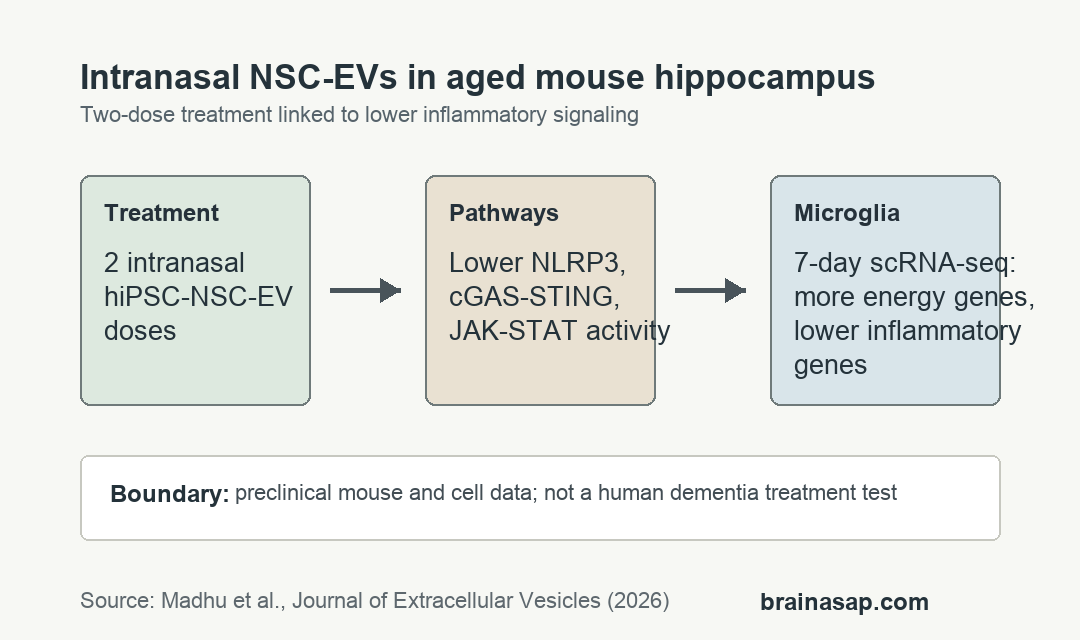
- Inflammation pathways fell: EV treatment reduced proteins linked to NLRP3 inflammasome, p38/MAPK, cGAS-STING-IFN-1, and JAK-STAT signaling.
- Two miRNAs were tested: Cell assays linked miRNA-30e-3p to NLRP3 inhibition and miRNA-181a-5p to STING-pathway inhibition.
- Seven-day microglia readout: Single-cell RNA sequencing 7 days after treatment found broad microglial transcriptome shifts toward oxidative phosphorylation and away from proinflammatory pathways.
Source: Journal of Extracellular Vesicles (2026) | Madhu et al.
Intranasal hiPSC-NSC-EVs Targeted Aging Microglia in the Hippocampus
Extracellular vesicles are small membrane-wrapped packets released by cells. Here, the vesicles came from human induced pluripotent stem cell-derived neural stem cells, shortened as hiPSC-NSC-EVs.
The study focused on neuroinflammaging, a chronic, low-grade inflammatory state that can build in the aging hippocampus. The hippocampus matters because it supports memory, object recognition, and flexible learning.
Rather than transplanting stem cells, researchers tested whether EVs could deliver regulatory cargo through the nose. The intranasal route is attractive in animal work because it can reach brain tissue without surgery.
- Animal model: late middle-aged male and female C57BL/6J mice beginning at 18 months of age.
- Treatment: two intranasal doses of hiPSC-NSC-EVs rather than repeated long-term dosing.
- Main tissue target: hippocampal microglia, the resident immune cells that can amplify inflammatory signaling in old brain tissue.
The work should be read as preclinical aging biology. It maps a possible route for lowering inflammatory activity in old brain tissue without testing a human anti-aging therapy.
NLRP3 and cGAS-STING Signals Decreased After EV Treatment
Aging microglia can activate multiple inflammatory systems at once. Madhu et al. emphasized the NLRP3 inflammasome, a protein complex that helps drive inflammatory cytokine release.
The other central pathway was cGAS-STING, a DNA-sensing system tied to type 1 interferon signaling. Both pathways are plausible contributors to chronic immune activation in old hippocampal tissue.
Compared with vehicle-treated aged mice, EV-treated mice showed lower hippocampal markers of astrocyte hypertrophy, microglial clusters, and oxidative stress. The primary paper also reported higher expression of antioxidant proteins and genes involved in mitochondrial respiratory-chain integrity.
The pathway pattern was broader than one marker. The reported set included NLRP3, p38/MAPK, cGAS-STING-IFN-1, and JAK-STAT activity.
- Inflammasome arm: NLRP3 and ASC inflammasome-complex activity was reduced after EV treatment.
- Interferon arm: proteins in the cGAS-STING-IFN-1 cascade were lower in treated mice.
- Mitochondrial arm: respiratory-chain gene expression shifted upward, supporting a less stressed cellular-energy profile.
miRNA-30e-3p and miRNA-181a-5p Helped Explain the Mechanism
The vesicles carried microRNAs, short regulatory RNA molecules that can tune gene expression. The study highlighted miRNA-30e-3p and miRNA-181a-5p because targeted depletion experiments linked them to separate inflammatory pathways.
In genetically engineered RAW-cell assays, EVs with and without selected microRNAs were tested against pathway activation. Removing specific microRNAs weakened the anti-inflammatory effect, supporting the idea that EV cargo was doing more than passively entering tissue.
- miRNA-30e-3p: linked to inhibition of the NLRP3 inflammasome pathway.
- miRNA-181a-5p: linked to inhibition of the STING pathway.
- Mechanistic value: depletion experiments make the cargo argument stronger than a simple before-after mouse comparison.
That mechanism is still preclinical. RAW-cell assays simplify biology, and mouse hippocampal microglia do not behave exactly like human aging brain tissue.
Single-Cell RNA Sequencing Found a Seven-Day Microglia Shift
The single-cell RNA sequencing piece matters because it looked inside microglia rather than treating the hippocampus as one mixed tissue sample. Researchers profiled microglia 7 days after treatment and reported widespread transcriptomic changes.
EV treatment increased expression of many genes tied to oxidative phosphorylation, the mitochondrial process cells use to generate energy. At the same time, expression dropped across abundant genes involved in inflammatory signaling.
Those patterns fit the protein data. Aged microglia appeared less locked into inflammatory pathway activation and more aligned with mitochondrial function after EV treatment.
The study also reported better cognitive and memory function in treated animals. Because the available abstract and source record do not provide every behavioral test value, the behavior finding belongs beside the anti-inflammatory and mitochondrial readouts rather than as a standalone effect-size claim.
The Mouse Data Are Promising but Not a Nasal Spray for Human Brain Aging
The clearest takeaway is narrow: two intranasal hiPSC-NSC-EV doses changed inflammatory and mitochondrial readouts in old mice. The study did not test whether a nasal spray can reverse human brain aging.
The practical interest is still real. A noninvasive delivery route, if validated, would be easier to imagine clinically than direct brain infusion or cell transplantation.
- Translation gap: human dose, delivery, durability, and safety remain unknown.
- Disease gap: the model studied aging hippocampus, not diagnosed Alzheimer’s disease or another human dementia.
- Evidence gap: the study supports pathway modulation, but it does not establish long-term clinical benefit.
For now, the work is best read as an aging-brain mechanism study. It suggests that EV cargo can reduce multiple inflammatory pathways in aged hippocampal immune cells, with cognitive readouts moving in the same direction.
Citation: DOI: 10.1002/jev2.70232. Madhu et al. Intranasal Human NSC-Derived EVs Therapy Can Restrain Inflammatory Microglial Transcriptome, and NLRP3 and cGAS-STING Signalling, in Aged Hippocampus. Journal of Extracellular Vesicles. 2026;15(2):e70232.
Study Design: Mouse aging intervention study with hippocampal protein assays, single-cell RNA sequencing, and in vitro microRNA-depletion assays.
Sample/Model: Late middle-aged 18-month-old male and female C57BL/6J mice treated with two intranasal hiPSC-NSC-EV doses and assessed at 20.5 months.
Key Statistic: Single-cell RNA sequencing was performed 7 days after treatment and showed broad microglial shifts toward oxidative phosphorylation and away from proinflammatory signaling.
Caveat: Preclinical mouse and cell data; human dosing, durability, safety, and dementia-treatment effects remain untested.