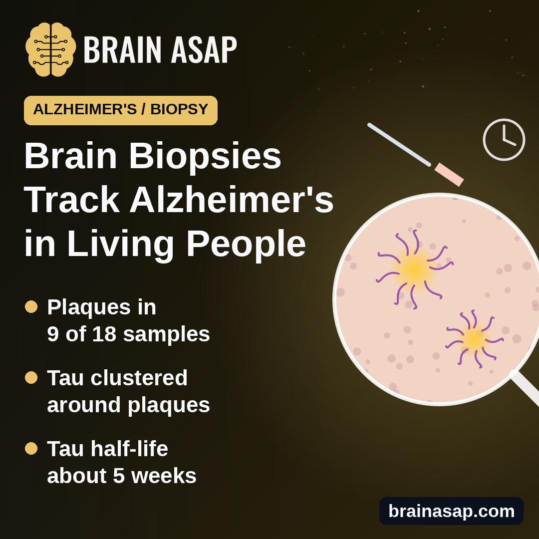
TL;DR: Tiny brain samples taken during routine shunt surgery let researchers study Alzheimer’s changes in living people. Half the samples had amyloid plaques, tau clustered around them, and brain tau had a half-life of about five weeks.
Key Findings
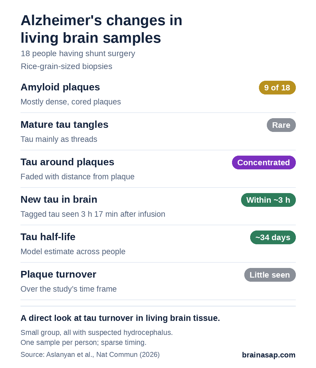
- 18 living patients: Rice-grain-sized samples taken during shunt surgery for suspected hydrocephalus.
- Plaques in 9 of 18: Mature tau tangles were rare.
- Tau gathered around plaques: Its signal faded with distance from each plaque.
- Brain makes tau fast: Labeled new tau showed up within about 3 hours.
- Tau half-life about 34 days: Plaques showed little turnover.
Source: Nature Communications (2026) | Aslanyan, Mukherjee et al.
Alzheimer’s disease starts changing the brain years before memory fails. But most of what we know about plaques and tangles comes from brains examined after death, or from indirect clues in spinal fluid, blood and scans.
A team led by researchers at University College London, Washington University in St. Louis and the University of Gothenburg found a way to look directly at living brain tissue.
A Grain of Brain From Routine Surgery
People with normal pressure hydrocephalus, a buildup of brain fluid that causes walking, bladder and thinking problems, often get a shunt to drain the fluid. Inserting the shunt tube already means passing through a small bit of brain. The team took a sample of that tissue, about the size of a grain of rice, from 18 patients.
The patients were older adults, mostly men, and all had some memory or thinking problems. None had complications from the biopsy.
Some patients also received an infusion of leucine, an amino acid tagged with a harmless heavy form of carbon, a few hours to months before surgery. Any protein made after the infusion carries the tag, so the team could “date” new tau in the tissue.
Plaques in Half, Tangles Rare
Nine of the 18 samples contained amyloid plaques, mostly dense, cored ones. Tau was present at low levels, mainly as thin threads and around plaques rather than as the mature tangles typical of later disease.
- Tau near plaques: A form of tau called pTau217 was concentrated in and around plaques and faded with distance.
- Spinal fluid mostly agreed: Fluid from the brain’s ventricles pointed to amyloid in 9 people; it matched the biopsy in 14 of 18.
- Two highest tau samples also had dense plaques.
Tau Is Made Fast and Lasts About Five Weeks
The tag showed up in brain tau as early as 3 hours and 17 minutes after the infusion started. By modeling tagged tau across patients sampled at different times, the team estimated a half-life of about 34 days for one tau fragment, similar to what has been measured in spinal fluid.
Plaque-containing samples tended to have more newly made tau, consistent with amyloid speeding tau production, though not every association held after statistical correction. Plaques themselves showed little sign of taking up new material over the study’s time frame.
A Small, Unusual Group
- Not typical Alzheimer’s patients: All had suspected hydrocephalus and symptoms.
- One small spot: Each biopsy sampled a tiny area of one brain region.
- Sparse timing: Each person gave one sample, so the half-life is a model estimate across people.
- Few controls: Comparisons used a handful of brains examined after death.
A New Window for Testing Treatments
Being able to measure how fast tau is made and cleared in living brain tissue could help show whether new Alzheimer’s drugs actually change these processes. The next step is larger groups and repeated measurements to confirm the five-week estimate and the link between plaques and new tau.
Citation: DOI: 10.1038/s41467-026-76494-4. Aslanyan A, Mukherjee S, Moncur EM, et al. Spatiotemporal characterization of Alzheimer disease pathology in living human brain tissue. Nat Commun. 2026;17:9878.
Study Design: Biopsy study with stable isotope labeling, mass spectrometry imaging and matched CSF, plus post-mortem comparators.
Sample Size: 18 living patients with suspected normal pressure hydrocephalus; 7 post-mortem brains.
Key Statistic: Model-derived brain tau (peptide 212–221) half-life 33.6 days.
Caveat: Small symptomatic surgical cohort; single-region biopsies; sparse timepoints.