
TL;DR: A 2026 systematic review in Molecular Psychiatry found that microglia-related findings across major depression, bipolar disorder, and schizophrenia were better explained by biological subgroups than by one uniform diagnosis-by-diagnosis inflammation pattern.
Key Findings
- Three evidence streams: The review integrated human translocator protein positron emission tomography (TSPO-PET) imaging, cerebrospinal fluid kynurenine-pathway metabolites, and postmortem microglial studies.
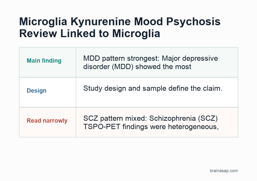
- MDD pattern strongest: Major depressive disorder (MDD) showed the most reproducible in vivo pattern, with increased TSPO binding in frontolimbic regions.
- SCZ pattern mixed: Schizophrenia (SCZ) TSPO-PET findings were heterogeneous, with small decreases or no change across studies.
- KYNA branch in SCZ: The most consistent biochemical finding in SCZ was a shift toward the kynurenic acid (KYNA) branch in CSF and cortex.
- BD data sparse: Bipolar disorder (BD) evidence was limited, including only one TSPO-PET study in the review’s summary.
Source: Molecular Psychiatry (2026) | Nussbaumer et al.
Microglia are the brain’s resident immune cells, and the kynurenine pathway is a tryptophan-metabolism route that can influence inflammation, glutamate signaling, and neural function. This review asked whether those systems show shared or different patterns across mood and psychotic disorders.
The review’s interpretation is not that one disorder is simply more inflamed than another. It argues for measurable subgroups and symptom dimensions that cut across diagnostic labels.
MDD Had the Clearest TSPO-PET Microglia Signal
Translocator protein positron emission tomography (TSPO-PET) is an imaging method often used as an indirect marker of neuroimmune activity. In the review, MDD showed the most reproducible in vivo pattern.
The strongest MDD pattern involved increased TSPO binding in frontolimbic regions. The review names the cingulate cortex, hippocampus, and prefrontal cortex as key areas.
- Cingulate cortex: A frontolimbic region involved in emotion, cognitive control, and conflict monitoring.
- Hippocampus: A memory and stress-sensitive structure often studied in depression.
- Prefrontal cortex: A region tied to executive control, emotion regulation, and illness burden.
Postmortem MDD findings were more cautious. At the diagnosis level, they were largely null for clear increases in microglial density or classical activation markers, though the review notes possible subtle homeostatic shifts.
That mismatch between imaging and postmortem evidence is one reason the review avoids a simple activation story.
TSPO-PET can reflect neuroimmune engagement in living patients, but it does not identify one cell type with perfect specificity. Postmortem studies also depend heavily on sampled brain region, illness stage, cause of death, and tissue handling.
The MDD finding is therefore best read as a reproducible imaging pattern, not as proof that all depression involves activated microglia. It may apply most strongly to subsets with stress exposure, inflammatory tone, illness severity, or treatment histories that align with frontolimbic immune engagement.
Schizophrenia Findings Pointed More Toward KYNA Than TSPO-PET
Schizophrenia did not show the same clean TSPO-PET pattern. The review describes SCZ imaging findings as heterogeneous, with small decreases or no change rather than one consistent increase.
The more consistent biochemical finding was a shift toward the kynurenic acid (KYNA) branch. Increased CSF KYNA and cortical KYNA fit with kynurenine-pathway effects on glutamatergic signaling.
- CSF KYNA: Cerebrospinal fluid findings suggested increased KYNA in schizophrenia.
- Cortical KYNA: Postmortem cortical findings also supported a KYNA-linked signal.
- Glutamate relevance: KYNA can affect NMDA-receptor signaling, a pathway often discussed in schizophrenia biology.
KYNA does not explain schizophrenia by itself. The narrower finding is that the review found a more reproducible pathway pattern there than a uniform microglial PET signature.
Bipolar Disorder Evidence Was Too Sparse for a Strong Pattern
BD was included descriptively because the available evidence was limited and heterogeneous. The review’s summary notes that TSPO-PET evidence for BD came from a single study reporting hippocampal increases.
Postmortem BD studies were also mostly unchanged at the diagnosis level. Subgroup analyses involving suicide or psychosis revealed findings that would be missed if all BD cases were treated as one biological category.
- Single PET study: The BD TSPO-PET base was too thin for firm imaging conclusions.
- Psychosis subgroup: Some kynurenine-pathway findings differed when psychotic features were considered.
- Suicide stratification: Postmortem findings could appear in clinically defined subgroups rather than in the whole diagnosis.
For psychiatric biomarkers, a diagnosis can contain several biological states, and averaging those states together can erase the measurable pattern.
Symptom Dimensions May Explain More Than Diagnostic Labels
The review’s interpretive claim is dimensional. Microglia-related findings may track inflammatory subtypes, suicidality, psychosis burden, cognitive impairment, illness stage, or treatment exposure more than DSM categories alone.
This helps explain why neuroinflammation studies can look inconsistent. Different samples may include different mixes of high-inflammation patients, chronic illness, acute episodes, medication exposure, and postmortem brain regions.
The review also separates kynurenine-pathway branches instead of treating the pathway as one marker. Kynurenine metabolism can move toward metabolites with different relationships to glutamate signaling, oxidative stress, and immune tone, so branch balance can matter as much as total pathway activity.
- Biological subgrouping: Future studies need inflammatory and clinical stratification before pooling patients.
- Longitudinal design: Repeated measures could show whether microglia-related findings change with episodes or remission.
- Better specificity: TSPO-PET has limited cellular specificity, so microglia-specific biomarkers remain a major need.
For psychiatric mechanism research, immune and microglial markers may be real, but the cleanest answers may come from symptom-linked subgroups rather than one marker for depression, bipolar disorder, or schizophrenia.
Clinically, that means biomarker studies need to record more than diagnosis. Psychosis severity, suicide history, cognitive impairment, episode phase, sleep disruption, medication exposure, and systemic inflammation could determine whether a microglia-related measure is visible or washed out.
Citation: DOI: 10.1038/s41380-026-03614-3. Nussbaumer et al. Multimodal microglial and kynurenine pathway alterations across the affective-psychosis spectrum: a systematic review of patterns, heterogeneity, and dimensional implications. Molecular Psychiatry. 2026.
Study Design: Systematic review integrating TSPO-PET, CSF kynurenine-pathway biomarkers, and postmortem studies.
Sample Size: Human literature review across MDD, BD, and SCZ, with quantitative synthesis focused on MDD versus SCZ.
Key Statistic: BD TSPO-PET evidence was limited to a single study, while MDD showed the most reproducible in vivo TSPO-PET pattern.
Caveat: TSPO-PET has limited cellular specificity, and many included studies had small samples or narrow brain-region coverage.