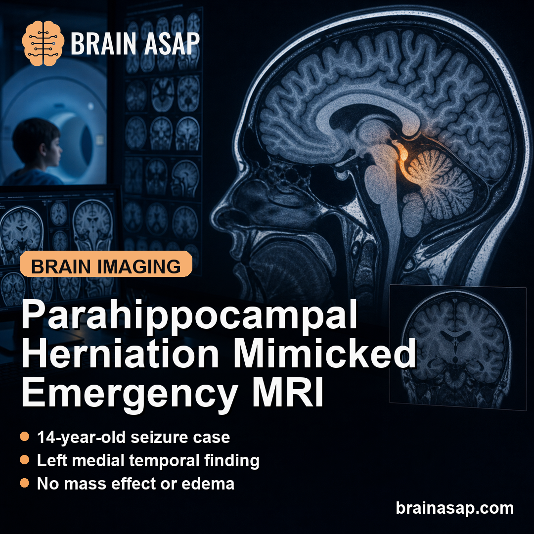
TL;DR: A 2026 study in Radiology Case Reports described a 14-year-old boy whose seizure workup found a rare congenital left parahippocampal herniation on MRI, with imaging features that favored a benign developmental variant rather than tumor or acute brain herniation.
Key Findings
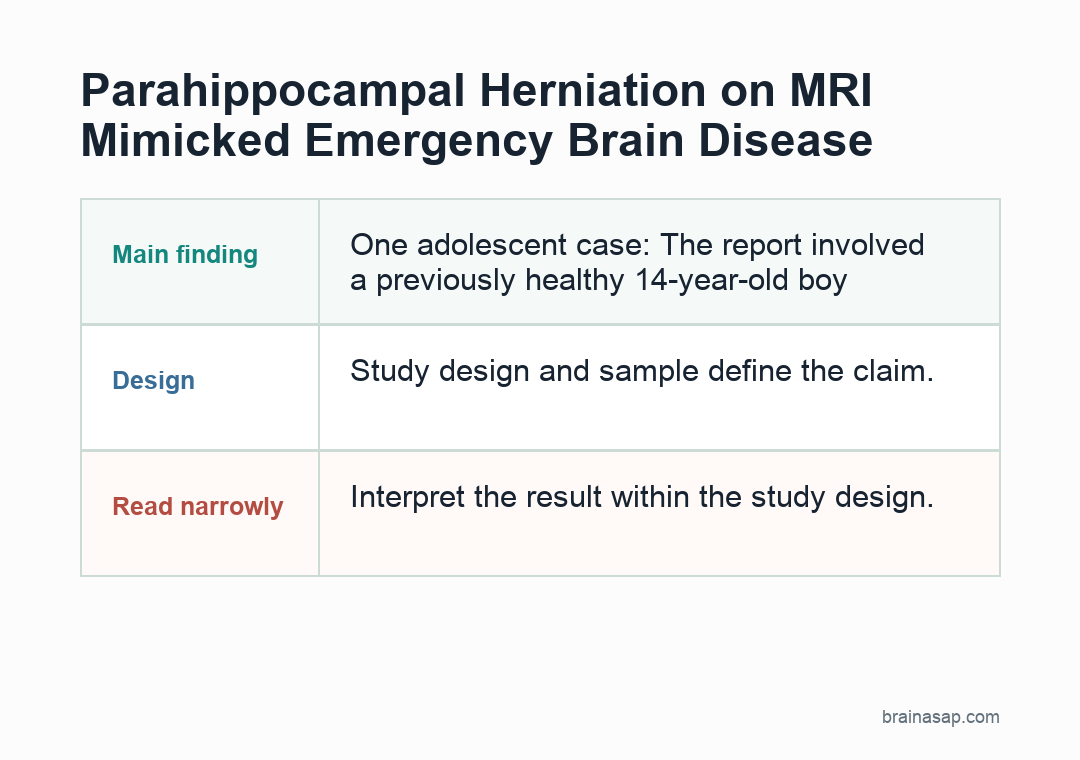
- One adolescent case: The report involved a previously healthy 14-year-old boy evaluated after a generalized tonic-clonic seizure lasting about 2 minutes.
- Normal initial workup: Neurological examination, laboratory tests, toxicology screening, and electroencephalography were reported as unremarkable.
- Left medial temporal finding: MRI showed left parahippocampal gyrus herniation through the tentorial incisura.
- No emergency features: Imaging showed preserved gray-white differentiation, no mass effect, no edema, no midline shift, no abnormal enhancement, and no restricted diffusion.
- Conservative course: The patient was discharged on carbamazepine 200 mg daily, but planned 3-month follow-up MRI was not completed.
Source: Radiology Case Reports (2026) | Elsheikh et al.
Congenital transtentorial parahippocampal herniation is an unusual MRI finding in which medial temporal-lobe tissue protrudes through the tentorial opening. The dangerous version of transtentorial herniation is usually caused by pressure from bleeding, tumor, swelling, trauma, or fluid buildup.
This report focused on the opposite problem: a stable-appearing congenital variant that can look alarming if the imaging context is missed. The case also shows why the word “herniation” needs clinical context; the same anatomical term can describe either a pressure emergency or a non-emergent developmental finding.
MRI Showed Left Parahippocampal Herniation Without Mass Effect
The patient presented after one generalized tonic-clonic seizure, followed by about 15 minutes of postictal confusion. There was no previous seizure history, no head trauma, no central nervous system infection, and no family history of epilepsy.
Brain MRI found a focal protrusion of the left parahippocampal gyrus through the tentorial incisura. The herniated tissue kept normal gray-white matter architecture.
- Structural stability: MRI showed no midline shift, hydrocephalus, cisternal compression, or brainstem compression.
- Tissue signal: Diffusion-weighted imaging and ADC maps showed no restricted diffusion, arguing against acute infarct-like injury.
- Contrast result: Post-contrast T1-weighted imaging showed no abnormal enhancement, reducing concern for an enhancing mass.
Those features fit a congenital or idiopathic herniation better than an emergency uncal herniation. They also helped separate the finding from a medial temporal lobe tumor.
Seizure Workup Did Not Prove the Lesion Caused Epilepsy
The seizure brought the patient to medical attention, but the report did not establish the herniation as the epileptic focus. The distinction is essential for interpreting a single case report.
The boy’s neurological examination was normal after recovery. Blood count, renal function, glucose, electrolytes, and toxicology testing were also within normal limits or negative.
Electroencephalography showed no epileptiform discharges. That does not rule out epilepsy, but it weakens a direct claim that the parahippocampal protrusion caused the seizure.
- Known event: A generalized seizure occurred and prompted MRI.
- Unproven link: The congenital herniation was detected during workup, not shown to generate seizures.
- Missing follow-up: No later seizure course or repeat imaging was available.
Benign Congenital Herniation Can Mimic Tumor or Emergency Herniation
Acute uncal herniation is a neurological emergency because rising intracranial pressure can compress the midbrain and cranial nerves. It can produce anisocoria, impaired consciousness, and brainstem dysfunction.
The congenital form described here lacked those warning signs. The patient was alert after the postictal period, hemodynamically stable, and neurologically intact.
Radiology recognition matters because a benign medial temporal protrusion can be mistaken for more serious pathology. The report specifically discussed the risk of confusing it with neoplasm, encephalocele, or pressure-related herniation.
Conservative Management Fit the Stable Imaging Findings
The patient received supportive acute care with a single intravenous diazepam dose. He was then discharged on carbamazepine 200 mg once daily.
For the imaging finding itself, the management logic was conservative. No surgery was described because there was no mass effect, edema, enhancement, hydrocephalus, or compression pattern suggesting a lesion that needed removal.
- Medication decision: Carbamazepine addressed seizure prevention after the clinical event.
- Imaging decision: Repeat MRI after 3 months was advised to check stability.
- Surgical decision: The MRI appearance supported watchful follow-up rather than invasive exploration.
That approach fits the conservative framing used in some prior descriptions of non-emergent descending transtentorial temporal-lobe herniations. In this setting, the imaging task is to rule out mass effect, abnormal enhancement, diffusion restriction, and pressure-related compression before considering invasive steps.
Follow-Up Limits Keep the Case Interpretation Narrow
The report’s largest limitation is follow-up. The planned repeat MRI could not be performed because of local instability, and no subsequent seizure or clinical follow-up data were available.
Repeat imaging would have been especially helpful because stability over time is one of the reassuring features of congenital herniation. Without it, the case rests mainly on the initial MRI appearance and the absence of acute neurological signs.
That leaves two separate statements. First, the MRI features were consistent with a benign congenital parahippocampal herniation. Second, the relationship between that finding and the seizure remained uncertain.
- What the case supports: A rare congenital medial temporal variant can appear during seizure imaging and mimic serious disease.
- What remains unresolved: The case does not show whether the herniation caused the seizure.
- Clinical caution: Stable imaging features should be matched with symptoms, EEG, and follow-up rather than interpreted in isolation.
For clinicians and families, the clinical value is recognition. When a neurologically stable patient has preserved tissue architecture and no pressure effects on MRI, congenital parahippocampal herniation belongs in the differential before assuming tumor or emergency herniation.
Citation: DOI: 10.1016/j.radcr.2026.02.017. Elsheikh et al. Congenital transtentorial parahippocampal herniation presenting with new-onset seizures in a 14-year-old boy. Radiology Case Reports. 2026;21:2449-2452.
Study Design: Single pediatric radiology case report.
Sample Size: One 14-year-old boy with new-onset generalized tonic-clonic seizure.
Key Statistic: MRI showed left parahippocampal herniation with no mass effect, edema, restricted diffusion, abnormal enhancement, hydrocephalus, or midline shift.
Caveat: Follow-up MRI and later seizure outcome data were unavailable, so causality between the imaging finding and seizure was not established.