TL;DR: A 2026 study in Signal Transduction and Targeted Therapy built a Parkinson’s visual hallucination mouse model and found that a combined behavior map identified hallucination-like episodes better than any single movement measure.
Key Findings
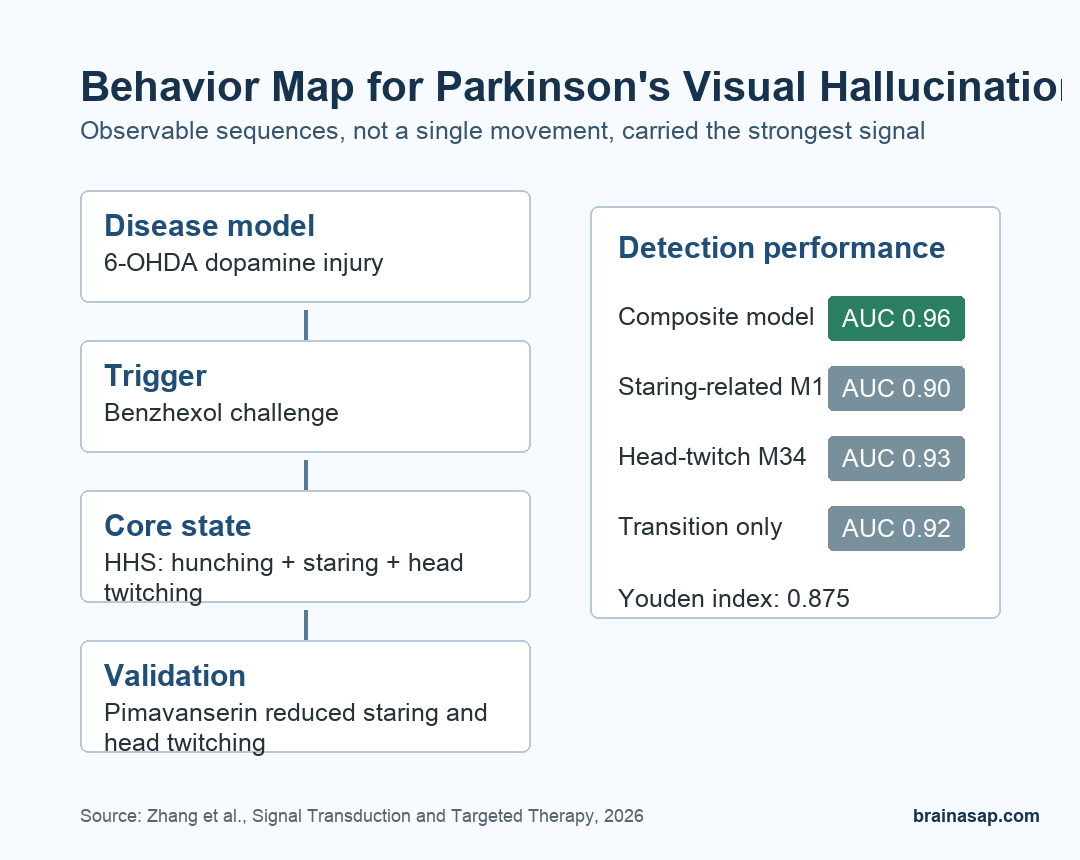
- Model: Researchers used 6-OHDA to create Parkinson-like dopamine injury in male C57BL/6 mice, then used benzhexol hydrochloride to trigger Parkinson’s visual hallucination-like behavior.
- Main behavioral state: The clearest marker was a hallucination-related hunching state, or HHS, combining sustained hunching, prolonged staring, and embedded head twitching.
- Classification: A composite model using transition patterns plus HHS reached an AUC of 0.96, compared with lower AUC values for single measures.
- Brain activity: PDVH mice showed increased c-Fos activation in the medial prefrontal cortex and primary visual cortex during staring and head-twitching episodes.
- Drug validation: Pimavanserin, a 5-HT2A receptor inverse agonist used for Parkinson’s psychosis, reduced staring and head-twitching durations in the model.
Source: Zhang et al. 2026.
Parkinson’s disease is usually recognized through movement symptoms, but many patients also develop neuropsychiatric symptoms. Visual hallucinations are one of the hardest to model because patients can describe experiences that laboratory animals cannot report directly.
That creates a measurement problem. Researchers cannot ask a mouse whether it saw a formed image.
They have to build a model where an internal state leaves a reliable behavioral trace. This study tested whether spontaneous movement sequences could serve that role for Parkinson’s visual hallucination research.
A Parkinson’s Mouse Model Was Paired With Benzhexol
Researchers first created a Parkinson-like mouse model by injecting 6-hydroxydopamine (6-OHDA) into the right dorsal striatum. This approach damages dopaminergic neurons, giving the study a disease-relevant background rather than a generic psychedelic or psychosis model.
After 4 weeks, the mice showed expected Parkinson-like deficits:
- Motor behavior: lower locomotor activity, shorter rotarod fall latency, longer pole descent time, and weaker grip strength.
- Dopamine injury: reduced tyrosine hydroxylase-positive cells in the substantia nigra on the lesioned side.
- Cognition: impaired novel object recognition and spatial memory performance.
The hallucination-like state was then induced with benzhexol hydrochloride, an anticholinergic drug used for Parkinsonian tremor that can produce hallucinations at excessive exposure. The paper calls the resulting condition PDVH, short for Parkinson’s disease-related visual hallucination.
Hunching, Staring, and Head Twitching Formed the Core Readout
The team used a temporal analysis of spontaneous behavior pipeline, or TASB, to organize movement into modules and transitions. The important result was not one isolated twitch.
It was a repeatable behavioral state that lasted long enough to analyze as a sequence.
The researchers named the combined posture-and-movement cluster the hallucination-related hunching state. HHS combined 3 observable features:
- Sustained hunching: a posture that anchored the behavioral state rather than a passing movement.
- Prolonged staring: a visual-attention-like component that matched the hallucination focus of the model.
- Embedded head twitching: a movement related to known hallucination-like readouts but placed inside a longer behavioral episode.
A single head-twitch response can be too narrow for this question. HHS gave researchers a broader readout that included posture, visual fixation, and timing.
Transition Patterns Improved Hallucination Detection
Researchers then tested whether the order of behaviors helped identify PDVH-like episodes. They built a spontaneous behavior transition map and found that the next behavior depended strongly on the immediately preceding state.
In other words, PDVH behavior was not random noise added to a Parkinson’s model. It had structure.
A composite classifier using transition patterns plus HHS performed best. The combined model reached an AUC of 0.96.
Single-feature approaches were lower: AUC 0.90 for one staring-related movement, AUC 0.93 for a head-twitching-related movement, and AUC 0.92 for transition features alone.
The authors also reported a Youden index of 0.875 for the integrated model, suggesting a better balance of sensitivity and specificity than isolated behavior measures.
For a laboratory model, that means the system can sort PDVH-like animals with less dependence on one fragile readout.
Brain Activity Supported the Behavioral Readout
The behavioral pattern was not the only evidence. During staring and head-twitching episodes, PDVH mice showed increased c-Fos activation in the medial prefrontal cortex and primary visual cortex.
c-Fos is commonly used as a marker of recent neuronal activity.
The brain regions are relevant because hallucinations involve more than the retina or the motor system. The model pointed toward altered visual and prefrontal activity, which fits the broader idea that Parkinson’s hallucinations involve abnormal visual processing, attention, and top-down interpretation.
The team also used single-nucleus RNA sequencing in the prefrontal cortex and midbrain. That molecular work was exploratory, but it helped connect the behavioral state with cell-level changes rather than leaving HHS as only an external movement label.
Pimavanserin Partly Normalized the Hallucination-Like State
The strongest validation step was pharmacological. Researchers treated PDVH mice with pimavanserin, a 5-HT2A receptor inverse agonist used clinically for Parkinson’s disease psychosis.
The treatment was given at 3 mg/kg/day on days 12 through 14, with the last injection 30 minutes before behavioral testing.
Pimavanserin significantly reduced the durations of staring and head twitching, while the overall transition frequency did not change in the same way. In a separate analysis, treated mice shifted toward an intermediate behavioral position between untreated PDVH mice and non-PDVH controls.
That result does not prove the model fully captures human hallucinations. It does show that the behavioral pattern responds to a drug tied to Parkinson’s psychosis, which makes the readout more useful for future mechanism and treatment studies.
The Caveat Is Translation From Behavior to Experience
This is a mouse study, and hallucinations are subjective experiences. The researchers are not showing that mice see what patients see.
They are showing that a Parkinson-like model plus anticholinergic challenge produces structured, drug-responsive behavior linked to visual and prefrontal activity.
Parkinson’s hallucination research may benefit from measuring behavioral sequences, not just isolated events.
If HHS and transition mapping hold up across laboratories, they could give researchers a more objective way to test circuits and candidate treatments before moving back to human studies.
Citation: DOI: 10.1038/s41392-026-02651-2. Zhang et al. Temporal assessment of behavior in Parkinson’s visual hallucinations via a multidimensional analysis strategy. Signal Transduction and Targeted Therapy. 2026.
Study Design: Parkinson’s disease mouse-model experiment with 6-OHDA lesions, benzhexol-induced hallucination-like behavior, temporal behavior mapping, brain-activity assays, sequencing, and pimavanserin validation.
Sample/Model: Male C57BL/6 mice; key behavior analyses included non-PDVH and PDVH groups with n = 16 per group, and pimavanserin validation included non-PDVH n = 15, PDVH n = 15, and PDVH + PIM n = 14.
Key Statistic: Composite transition-plus-HHS classification reached AUC = 0.96, outperforming single behavioral parameters.
Caveat: The model measures observable hallucination-like behavior in mice, not subjective visual experience in people with Parkinson’s disease.