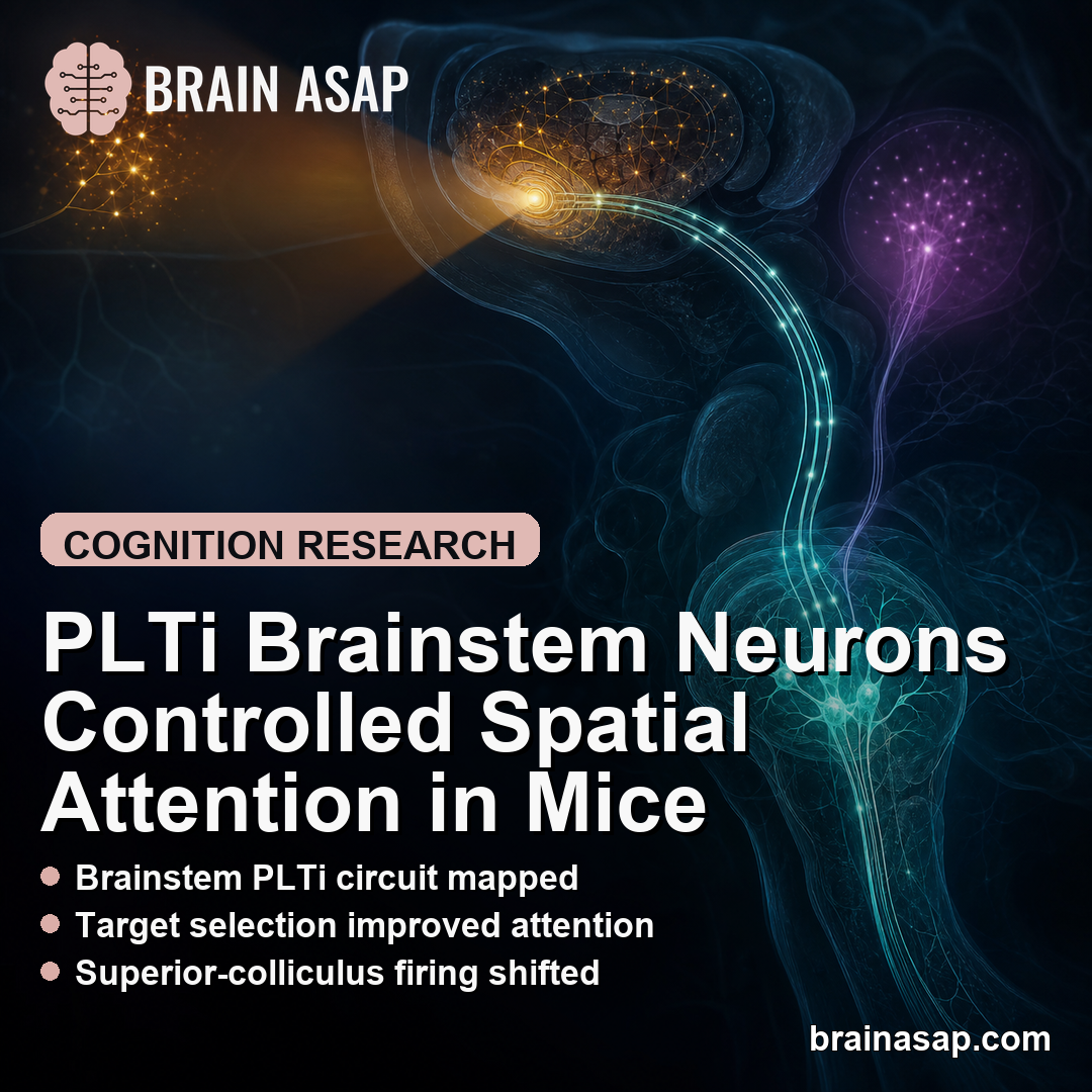
TL;DR: A 2026 study in Nature Communications found that PLTi, an evolutionarily old group of inhibitory brainstem neurons, was required for mice to choose a goal-relevant visual target over lower-priority distractors.
Key Findings
- PLTi defined: Parabigemino-lateral tegmental inhibitory complex neurons were mostly parvalbumin-positive GABAergic cells in the mouse brainstem.
- 95.01% overlap: Nearly all GABA-positive cells in the parabigemino-tegmental complex also labeled for parvalbumin.
- 10-task mice: Freely moving mice performed a touchscreen flanker task that separated target selection from simple sensory detection.
- Silencing deficit: Chemogenetic PLTi silencing impaired incongruent target selection while leaving congruent trials and single-target control behavior intact.
- SC pathway: PLTi shaped neural competition in the intermediate and deep superior colliculus, a midbrain attentional hub.
Source: Nature Communications (2026) | Kothari et al.
PLTi neurons sit in a brainstem region that resembles older vertebrate attention circuits. The study asked whether this inhibitory cell group helps the brain pick the behaviorally relevant location when another stimulus competes for control.
The clearest deficit appeared in conflict trials. When the distractor pointed against the target choice, silencing PLTi made mice more likely to choose incorrectly without producing a broad sensory or motor failure.
PLTi Neurons Connected Brainstem Inhibition to the Superior Colliculus
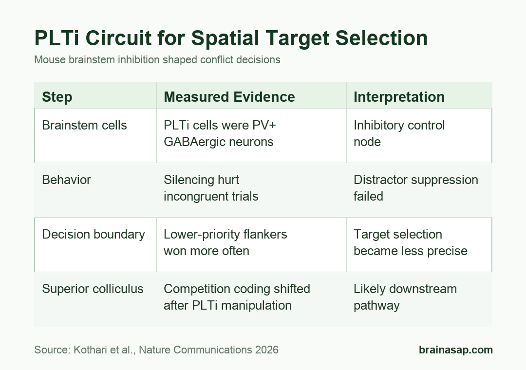
The researchers first identified PLTi anatomically and functionally. Immunostaining showed that the relevant parabigemino-tegmental complex cells were overwhelmingly inhibitory and parvalbumin-positive.
The study reported 95.01% +/- 0.46% of GABA-positive cells also labeled for parvalbumin, while 94.33% +/- 1.33% of parvalbumin-positive cells also labeled for GABA.
- Cell type: PLTi cells were parvalbumin-positive GABAergic neurons.
- Projection target: Viral tracing showed projections to the intermediate and deep superior colliculus.
- Functional effect: Chemogenetic activation of PLTi suppressed spontaneous activity in superior-colliculus neurons.
Activation data made the circuit direction clearer. Superior-colliculus firing dropped from 47.01 spikes/s at baseline to 7.2 spikes/s 30 minutes after CNO and 5.9 spikes/s 60 minutes after CNO.
Those measurements support an inhibitory control pathway: PLTi can change which competing spatial representation wins inside the superior colliculus.
PLTi Silencing Impaired Incongruent Target Selection
The behavioral test was a mouse touchscreen flanker task. A target appeared with or without flankers, and mice had to choose the target-defined response rather than follow distracting stimuli.
The task distinguished congruent trials, where target and flanker supported the same response, from incongruent trials, where a distractor pulled behavior toward the wrong choice.
- Single target: Mice could respond to target orientation without a competing flanker.
- Congruent flanker: Target and flanker supported the same response direction.
- Incongruent flanker: Target and flanker supported opposite responses, creating the attention-control demand.
Bilateral PLTi silencing selectively hurt performance in the incongruent condition. Congruent flanker trials remained intact, and a control group that received CNO without the inhibitory receptor did not show the same deficit.
The deficit also appeared with weak incongruent flankers. A simple salience explanation would not predict that lower-priority distractors become more behaviorally disruptive after PLTi silencing.
Target-Selection Errors Were Not Explained by Basic Perception or Movement
A key risk in attention studies is mistaking a broad sensory or motor problem for an attention-specific problem. The paper addressed that by testing several control explanations.
- Single-stimulus perception: PLTi silencing did not impair performance in a single-target discrimination task.
- Movement selection: The manipulation did not create a general task-relevant motor-plan selection problem.
- Response time: Reaction times became faster, but accuracy dropped only in incongruent trials, which did not match a simple speed-accuracy tradeoff.
The trial-by-trial profile points to a specific function. PLTi was needed when the animal had to use goal relevance to suppress a competing location.
The reaction-time result made the control logic stronger. If the manipulation only made mice respond too quickly, accuracy should have fallen across the task, but the error increase was concentrated in the condition where the target and flanker competed.
In the paper’s drift-diffusion modeling, PLTi silencing affected the decision boundary separating the target from the flanker. Behavior became less categorical, and lower-priority flankers more often outcompeted the target.
Superior-Colliculus Recordings Linked PLTi to Neural Competition
The superior colliculus is already known as a spatial orienting and attention hub. This study added a candidate inhibitory input that may help sculpt winner-take-all-like spatial decisions.
In passive head-fixed mice, the researchers recorded from the intermediate and deep superior colliculus during competing-stimulus presentations. PLTi manipulation changed how superior-colliculus neurons represented the stronger versus weaker stimulus.
- Behavioral boundary: Silencing PLTi shifted the target-versus-flanker decision boundary.
- Neural boundary: Superior-colliculus representation of competing stimuli also became less sharply separated.
- Mechanistic pathway: Brainstem inhibition may help the superior colliculus suppress lower-priority distractor representations.
This is a mouse circuit study, not direct evidence that the same named cell group controls human attention. The evolutionary argument comes from conserved circuit anatomy and the similarity to older vertebrate inhibitory attention systems.
Mouse Chemogenetic Data Limit the PLTi Attention Claim
The main limitation is translation. Freely moving mice can model target selection, but the experiment does not measure human attention disorders, conscious focus, or clinical distractibility.
Experiments and analyses were not blinded, and the paper states that no power analysis predetermined sample size. The sample sizes were described as similar to field norms, but that is not the same as a prospective power plan.
Within those limits, the paper identifies a concrete circuit role: PLTi helped keep a goal-relevant visual target in control when a lower-priority distractor competed for the response.
Citation: DOI: 10.1038/s41467-026-72340-9. Kothari et al. Evolutionarily old brainstem neurons are required for the control of selective spatial attention. Nature Communications. 2026;17:5849.
Study Design: Mouse circuit-neuroscience study using anatomy, chemogenetics, touchscreen behavior, modeling, and superior-colliculus recordings.
Sample/Model: Freely moving and head-fixed mice, including 10 mice in the main flanker-task performance comparison.
Key Statistic: PLTi activation reduced superior-colliculus firing from 47.01 spikes/s at baseline to 7.2 spikes/s after 30 minutes and 5.9 spikes/s after 60 minutes.
Caveat: The evidence is from mouse circuit manipulation and does not directly establish the same mechanism in human attention.