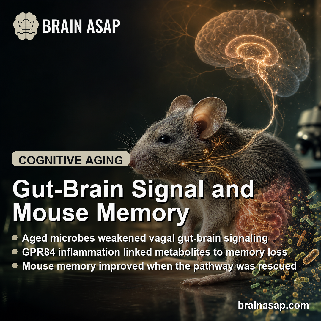
TL;DR: A 2026 mouse study in Nature found that age-related gut microbiome changes impaired vagal gut-brain signaling and hippocampal memory encoding, while several interventions restored memory performance in aged or microbiome-aged mice.
Key Findings
- Aged microbiome transfer impaired memory: Young mice exposed to aged gut microbes showed worse novel object recognition and Barnes maze performance without a matching drop in general exploration.
- Parabacteroides goldsteinii emerged as a driver: The bacterium increased with age, transferred through co-housing or microbiota transplantation, and caused memory impairment when given to young mice.
- Vagal gut-brain signaling weakened: Aged microbiome exposure reduced nutrient-evoked activation of vagal sensory neurons and blunted hippocampal FOS responses during memory tasks.
- Medium-chain fatty acids acted through GPR84: Microbial metabolites such as 3-HOA and decanoic acid triggered GPR84-dependent peripheral myeloid inflammation linked to memory loss.
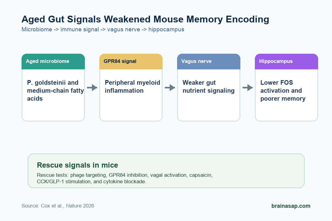
- Several rescue strategies worked in mice: Phage targeting of P. goldsteinii, GPR84 inhibition, vagal stimulation, capsaicin, CCK, GLP-1 stimulation, and inflammatory-cytokine blockade improved memory readouts in specific experiments.
The paper does not say the human gut microbiome already explains age-related cognitive decline. It maps a detailed mouse mechanism: aged gut bacteria changed peripheral immune signaling, weakened vagal interoception, and left the hippocampus less able to encode new information.
The mechanism turns a broad gut-brain idea into a testable circuit. The pathway runs from Parabacteroides goldsteinii and microbial fatty acids to GPR84 signaling in peripheral myeloid cells.
From there, the pathway moves to vagal sensory dysfunction, weaker hippocampal FOS activation, and poorer memory-task performance.
Aged Gut Microbes Were Enough to Impair Mouse Memory
Researchers first asked whether an aged microbiome could move memory performance in otherwise young mice. Co-housing young mice with aged mice shifted the young animals toward an old-like microbial state and impaired short-term memory in the novel object recognition task.
The same pattern appeared in spatial learning and memory on the Barnes maze.
The team then separated microbiome transfer from social exposure. Young germ-free mice given fecal microbiota from aged donors developed memory deficits.
By contrast, young germ-free mice co-housed with old germ-free mice kept normal cognitive performance. Antibiotic depletion of the aged microbiome also protected young mice from the co-housing effect.
Several control results were important:
- Young-young co-housing: Housing young mice with other young mice did not reproduce the memory deficit.
- Exploration behavior: The memory-task drop was not explained by reduced exploratory behavior.
- Germ-free aging: Germ-free mice showed delayed cognitive decline compared with conventional mice.
- Post-deficit antibiotics: 2 weeks of antibiotic treatment after impairment improved memory performance in microbiome-aged young mice.
Together, those experiments support a causal microbiome contribution in mice rather than a simple correlation between old age and worse cognition.
Parabacteroides Goldsteinii Became the Main Bacterial Suspect
The study charted microbiome aging across the mouse lifespan and found 1,133 species with significant age-related abundance changes. Researchers then ranked taxa by whether they increased with age, transferred through microbiome-aging experiments, and plausibly affected cognition.
Parabacteroides goldsteinii ranked as the top candidate. Its abundance rose with age, and stool proteomics confirmed increased Parabacteroides peptides.
Colonizing germ-free or antibiotic-treated mice with Parabacteroides goldsteinii caused memory impairment. Young mice from a facility with naturally high Parabacteroides goldsteinii also showed reduced memory function.
Other tested bacteria did not show the same effect. The contrast argues against a generic “old microbiome is bad” interpretation.
The measured pathway came from specific bacterial and metabolic features that changed gut-brain communication.
Vagal Interoception Connected the Gut Signal to the Hippocampus
The hippocampus was the central brain readout. The aged microbiome did not reproduce every hallmark of brain aging in young mice.
Hippocampal neurogenesis, astrogliosis, and dendritic spine structure did not explain the transferred memory deficit. Instead, the clearest change was weaker activity-dependent hippocampal activation during novelty exposure.
Researchers then focused on interoception, the nervous system’s sensing of internal body conditions. In young mice carrying an aged microbiome, gut nutrient infusion produced weaker activation in vagal sensory neurons.
Manipulating those neurons changed memory performance.
The vagal-signaling evidence included several converging tests:
- TRPV1-positive sensory neurons: Removing or silencing these neurons reduced hippocampal activation and impaired novel object recognition.
- PHOX2B-positive vagal neurons: Silencing vagal, rather than spinal, sensory populations reproduced the cognitive deficit.
- Vagal activation: Chemogenetic activation, low-dose capsaicin, CCK, GLP-1, and liraglutide restored memory readouts in specific aged or microbiome-aged mouse experiments.
This is the study’s most clinically relevant idea, but also its main boundary. The interventions show that stimulating gut-brain signaling can rescue mouse memory tasks under these conditions.
They do not establish a human anti-dementia treatment.
GPR84 Myeloid Inflammation Explained the Metabolite Step
The mechanism narrowed further when researchers examined microbial metabolites. Parabacteroides goldsteinii produced medium-chain fatty acids, including 3-HOA and decanoic acid. These compounds impaired memory performance and reduced hippocampal FOS responses in mice.
The key receptor was GPR84, which is largely expressed by myeloid immune cells. GPR84-deficient mice were protected from medium-chain-fatty-acid effects.
The GPR84 inhibitor PBI-4050 also restored memory performance in aged mice and in mice exposed to P. goldsteinii.
The immune experiments pointed outside the brain. Depleting peripheral myeloid cells, disrupting CCR2-dependent peripheral recruitment, or removing GPR84 from the hematopoietic compartment protected cognition.
TNF and IL-1beta signaling also mattered, and cytokine blockade improved memory readouts in aged or microbiome-aged mice.
The Mouse Circuit Map Still Needs Human Testing
The strongest supported claim is precise: in mice, age-associated gut microbiome changes can impair memory by weakening vagal interoceptive signaling to the hippocampus through a P. goldsteinii – medium-chain fatty acid – GPR84 – myeloid inflammation pathway.
That boundary should prevent overreach. The study does not show that taking capsaicin, GLP-1 drugs, antibiotics, probiotics, or anti-inflammatory agents will improve memory in older adults.
It also does not show that a single bacterium explains human cognitive aging. Mouse microbiomes, controlled co-housing, germ-free systems, and engineered vagal manipulations are powerful experimental tools, but they are not everyday human aging.
The study still gives cognitive-aging research a more concrete target. Instead of treating the gut-brain axis as a vague wellness category, it identifies bacterial taxa, metabolites, immune receptors, sensory neurons, and hippocampal activation as linked steps in the same pathway.
Future human work can now test those steps directly instead of leaning on broad microbiome language.
Citation: DOI: 10.1038/s41586-026-10191-6. Cox et al. Intestinal interoceptive dysfunction drives age-associated cognitive decline. Nature. 2026;652:442-450.
Study Design: Mouse microbiome-aging study using co-housing, fecal microbiota transfer, germ-free and antibiotic experiments, bacterial colonization, metabolite testing, vagal-neuron manipulation, immune-cell experiments, and hippocampal activity readouts.
Sample/Model: Young and aged mice across multiple strains and facilities, including germ-free mice, genetically modified sensory-neuron and immune-signaling models, and microbiome/metagenomic lifespan mapping.
Key Statistic: Lifespan microbiome mapping found 1,133 age-altered species, with P. goldsteinii ranked as the top candidate bacterial driver of the mouse memory phenotype.
Caveat: The evidence is mechanistic and preclinical; human cognitive-aging prevention or treatment would need direct clinical testing.