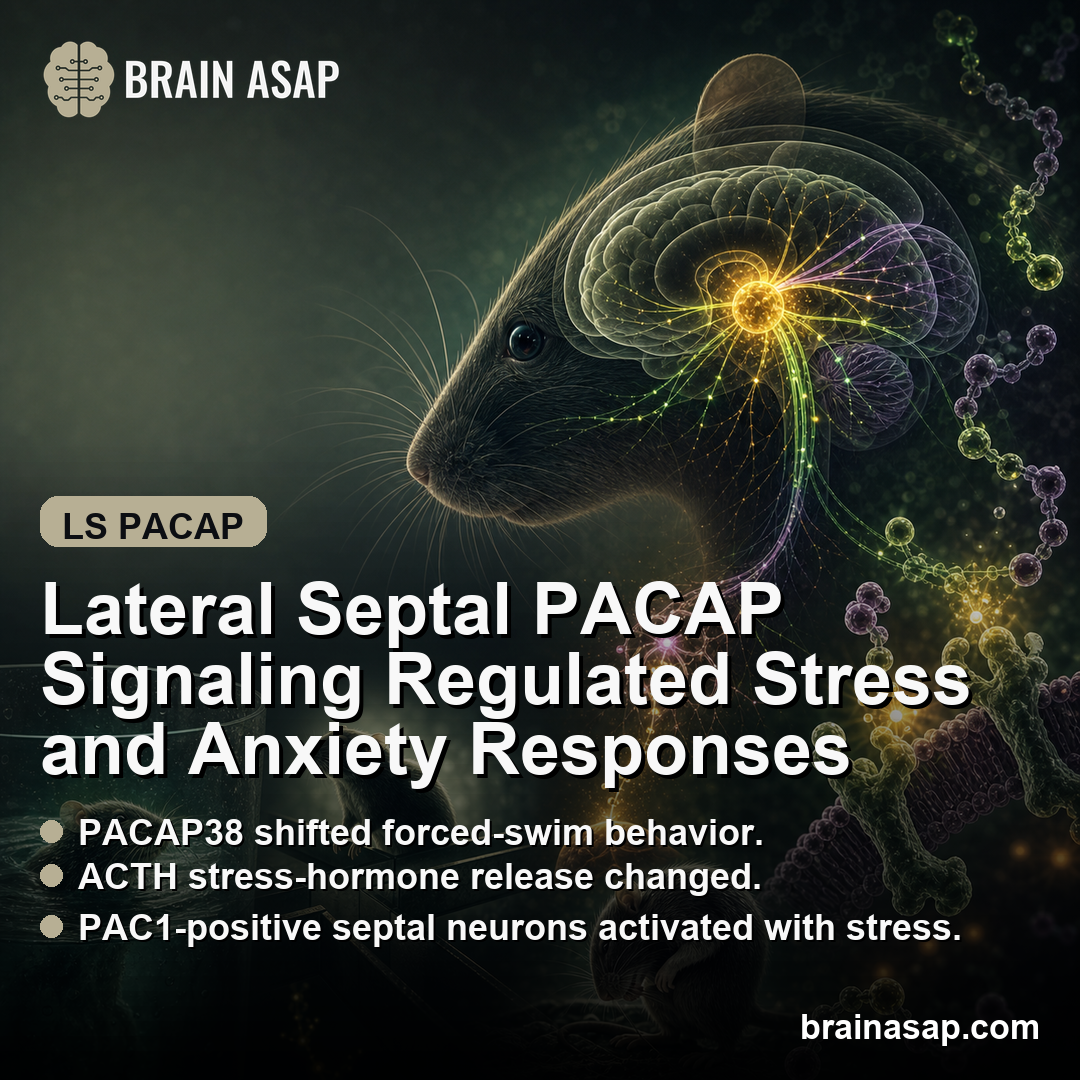
TL;DR: A 2026 rat study in Neuropsychopharmacology found that PACAP/PAC1 signaling in the lateral septum changed forced-swim coping, ACTH stress-hormone release, open-arm exploration, and grooming behavior.
Key Findings
- Stress changed PACAP signals by brain region: Acute swim stress increased PACAP mRNA in the lateral septum, BNST, and basolateral amygdala, while chronic variable mild stress reduced lateral-septum PACAP mRNA by 35%.
- PACAP38 shifted forced-swim behavior: Infusing PACAP38 into the lateral septum increased floating and reduced struggling, especially at the higher 150 pmol/site dose.
- ACTH rose during stress, not at baseline: Lateral-septum PACAP38 increased ACTH after forced swim stress at 10 and 30 minutes, but did not produce the same sustained hormone rise without stress.
- The elevated plus maze pointed in the same direction: PACAP38 reduced open-arm entries and open-arm time, while PACAP(6-38) blockade increased open-arm exploration.
- The finding is still preclinical: These were male rat experiments using local brain infusions, not a human PTSD, depression, or anxiety treatment study.
Source: Neuropsychopharmacology (2026) | Fontebasso et al.
PACAP stands for pituitary adenylate cyclase-activating polypeptide, a neuropeptide involved in stress signaling. PAC1 is one of the main receptors that can respond to PACAP.
This Neuropsychopharmacology paper asked a specific circuit question: does PACAP/PAC1 signaling inside the lateral septum help control stress-coping behavior, ACTH hormone release, open-arm exploration, and grooming in rats?
The study tracked four connected readouts:
- Gene expression: PACAP and PAC1 receptor mRNA after stress exposure.
- Behavior: forced-swim coping, open-arm exploration, and grooming.
- Hormone output: ACTH after forced swim stress.
- Cell activation: PAC1 and c-Fos staining in lateral septum neurons.
The lateral septum is a limbic region connected with hippocampal, hypothalamic, and brainstem-related stress pathways. Those connections make it a plausible place for stress signals to affect both behavior and hormone output.
PACAP and PAC1 Were Measured Across Limbic Stress Circuits
Measurement detail: The researchers measured PACAP and PAC1 receptor mRNA after repeated forced swim stress and after 7 days of chronic variable mild stress.
Acute swim stress increased PACAP mRNA in the lateral septum, BNST, and basolateral amygdala. It did not produce the same PACAP change in the PVN, central amygdala, or medial amygdala.
Chronic variable mild stress produced a different pattern. Lateral-septum PACAP mRNA fell by 35%, while PAC1 receptor expression increased in the BNST.
Takeaway: PACAP was not just a generic stress marker. The direction of change depended on the stress model, the brain region, and whether the researchers measured PACAP itself or the PAC1 receptor.
Lateral Septum PACAP38 Shifted Forced-Swim Behavior and ACTH
The main manipulation was local PACAP38 infusion into the lateral septum before forced swim testing in male rats.
At 150 pmol/site, PACAP38 increased floating and reduced struggling. In this assay, floating is treated as passive coping, while struggling reflects more active escape-like behavior.
The hormone result was not a simple baseline effect.
PACAP38 increased ACTH after forced swim stress, especially at 10 minutes and 30 minutes, but did not create the same sustained ACTH rise without the swim stress challenge.
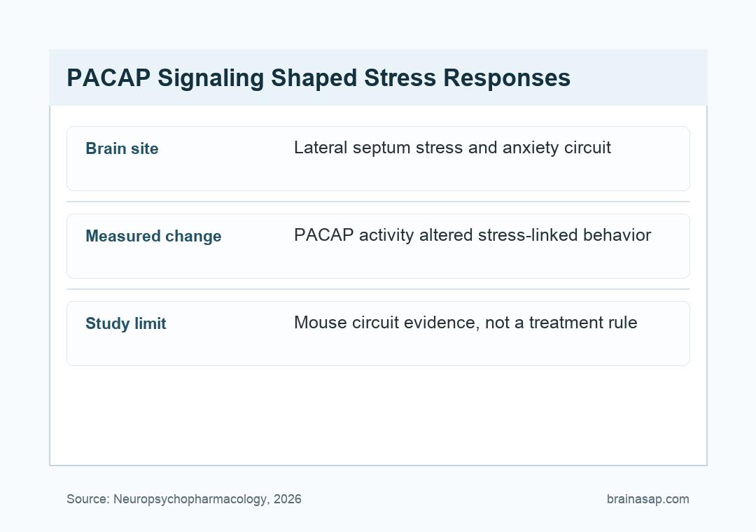
Control detail: PACAP38 infusion into the rostral anterolateral BNST did not significantly change forced-swim behavior or ACTH. That control makes the lateral septum claim more specific.
PACAP38 Changed Open-Arm Exploration and Grooming
In the elevated plus maze, lateral-septum PACAP38 reduced open-arm exploration. The study reported an almost 30% reduction in open-arm entries and about a 50% decrease in open-arm time.
Those changes were not explained by lower movement, because closed-arm entries and total distance traveled did not significantly differ between groups.
The antagonist result strengthened the interpretation. PACAP(6-38), a PACAP receptor antagonist, increased open-arm entries and open-arm time when infused into the lateral septum.
The same PACAP38-treated rats were also tested in the sucrose splash test. PACAP38 reduced grooming time and cut grooming bouts by almost 50%, while latency to first grooming did not significantly change.
Takeaway: The lateral septum PACAP result was not based on one behavioral readout. It appeared across forced-swim coping, ACTH release, elevated-plus-maze exploration, and sucrose-splash grooming.
PAC1-Positive Lateral Septum Neurons Were Activated by Stress
The circuit evidence came from PAC1 and c-Fos staining after forced swim stress. c-Fos marks recently activated neurons, and PAC1 marks neurons positioned to respond to PACAP.
The study found PAC1 receptor-expressing neurons across the lateral septum, with PAC1 staining on the membrane and proximal dendrites of many c-Fos-positive neurons. The overlap appeared more prominent in the ventral lateral septum.
Researchers point to possible downstream GABAergic pathways from the lateral septum to regions such as the dorsal periaqueductal gray, lateral hypothalamus, anterior hypothalamic area, PVN, and PAG-linked stress circuits.
Takeaway: The study supports lateral-septum PACAP/PAC1 signaling as a stress-circuit mechanism, but it does not yet identify the exact PAC1-positive cell type or projection that drives each behavioral output.
Male Rat PACAP Findings Do Not Equal Human Treatment Claims
Directly supported: In male rats, lateral-septum PACAP/PAC1 signaling changed forced-swim behavior, ACTH release during stress, elevated-plus-maze behavior, and grooming.
Plausible: The lateral septum may be one place where PACAP-sensitive neurons translate stress exposure into a coordinated behavioral and endocrine response.
Speculative: A similar pathway could matter for human stress-related conditions, but this study did not test humans, symptoms, medications, psychotherapy, or clinical outcomes.
- Species: The experiments were done in rats.
- Sex: The experiments used male rats, so female stress biology remains a major open question.
- Method: Local microinjection is helpful for circuit testing, but it is not the same as a selective clinical intervention.
- Receptor specificity: PAC1 is the most plausible receptor in the lateral septum, but more selective tools would make the receptor claim cleaner.
The next direct test is not simply whether PACAP is “important” for stress. It is which lateral-septum PAC1-positive neurons, which projections, and which stress conditions drive each part of the phenotype.
Final takeaway: This is good preclinical circuit evidence for PACAP/PAC1 signaling in the lateral septum.
It should be read as a rat stress-biology finding with clear behavioral and hormone endpoints, not as a ready-made explanation for human anxiety, PTSD, or depression.
Citation: DOI: 10.1038/s41386-026-02409-y. Fontebasso et al. Lateral septal PACAP signaling regulates stress and anxiety reactions. Neuropsychopharmacology. 2026.
Study Design: Preclinical rat study using acute forced swim stress, chronic variable mild stress, qPCR, intra-lateral-septum PACAP38/PACAP(6-38) microinjection, ACTH sampling, elevated plus maze, sucrose splash testing, and PAC1/c-Fos immunohistochemistry.
Sample Size: Adult male Sprague-Dawley rats; key behavioral cohorts included intra-LS PACAP38 forced-swim groups of n=5-8, ACTH groups of n=6 per group, elevated-plus-maze groups of n=7-9, and PACAP(6-38) groups of n=7-9.
Key Statistic: PACAP38 in the lateral septum increased passive floating (F(2,17)=8.51, p=0.0027), increased ACTH AUC (t(10)=3.466, p=0.0061), reduced open-arm time by about 50%, and PACAP(6-38) increased open-arm exploration.
Caveat: Male rat circuit experiments using local microinjection; the study does not test human anxiety, PTSD, depression, or treatment outcomes.