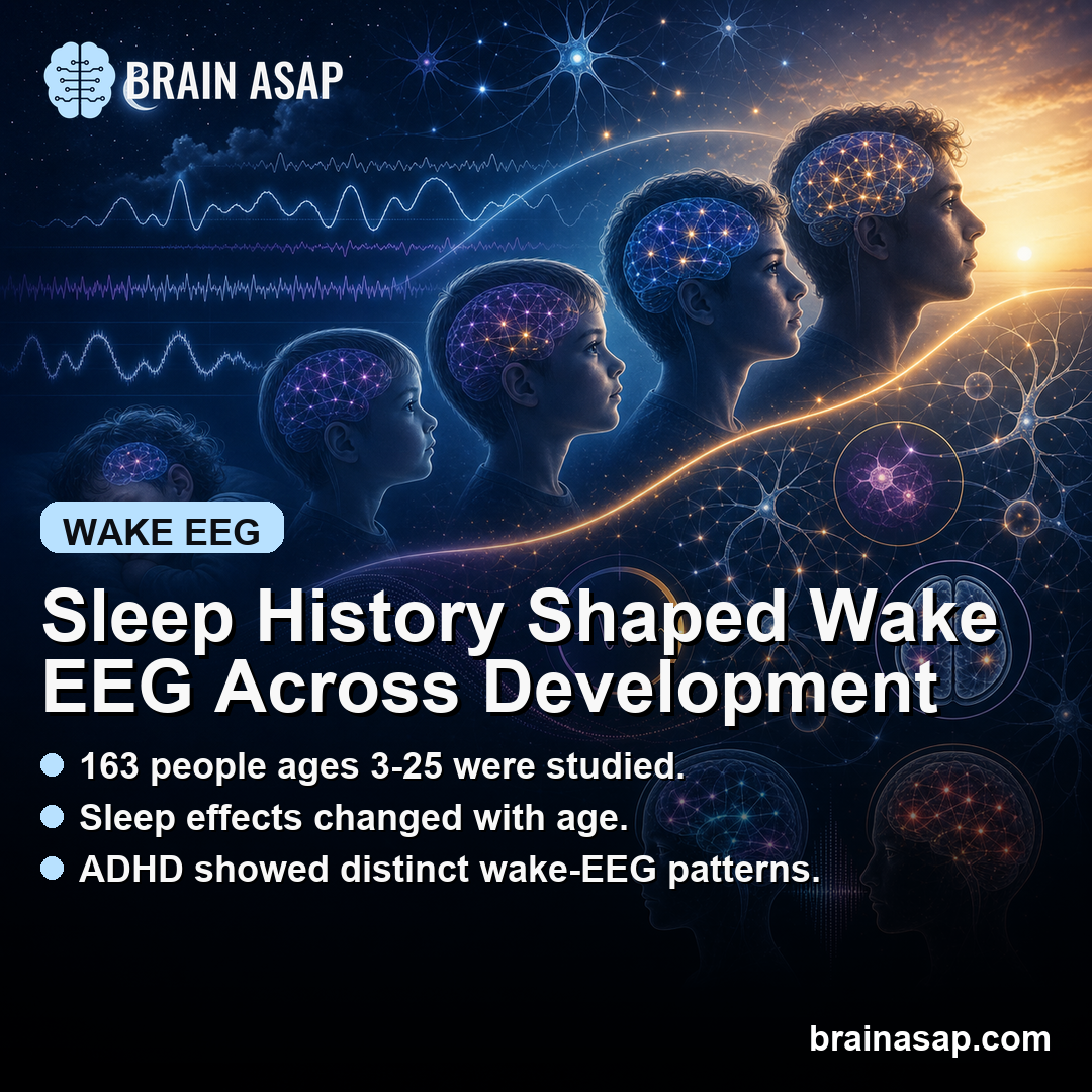
TL;DR: A 2026 eNeuro study of 163 people ages 3-25 found that sleep history and age shaped waking electroencephalography (EEG) oscillations, while children with ADHD showed distinct sleep-wake EEG patterns.
Key Findings
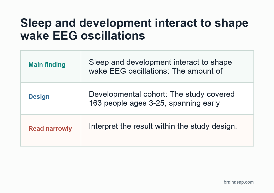
- Sleep and development interact to shape wake EEG oscillations: The amount of recent sleep or wakefulness produces age-dependent effects on EEG oscillation amplitude and density during waking, not just during sleep.
- Developmental cohort: The study covered 163 people ages 3-25, spanning early childhood through young adulthood.
- Both periodic and aperiodic activity affected: Researchers analyzed both oscillation amplitudes and density (periodic activity) and aperiodic activity (offsets and exponents) — the broadband background EEG activity that doesn’t fit into oscillation peaks.
- ADHD showed distinct patterns: Comparison with children diagnosed with ADHD revealed different sleep-wake EEG dynamics, linking ADHD-relevant physiology to sleep history.
- Sex representation: The cohort included 62 female participants, which matters because sleep and EEG maturation can differ by sex.
Source: eNeuro (2026) | Snipes et al.
Electroencephalography, or EEG, records the brain’s electrical activity from the scalp. Sleep research has long shown that sleep slow waves track recent wakefulness.
More time awake increases sleep pressure and generates more slow-wave activity in later non-REM sleep. The newer question is whether the same sleep-and-development interaction shapes waking EEG oscillations, the activity patterns recorded during ordinary wake periods.
Sleep History Can Shape Wake EEG
The two-process model of sleep regulation has dominated sleep neuroscience for decades. It posits that sleep is regulated by:
- Process S: Homeostatic sleep pressure that builds with wakefulness and dissipates during sleep, indexed by EEG slow-wave activity during NREM sleep.
- Process C: Circadian rhythm signaling that determines preferred timing of sleep and wake.
Most research on Process S has focused on how it shapes sleep EEG, especially slow-wave activity during non-REM sleep.
Sleep pressure also affects waking brain function. Tired people have measurably different cognitive performance, attention, and arousal than rested people, so wake EEG oscillations give researchers a way to quantify those differences.
Researchers tested the sleep-development link systematically across development.
Wake EEG Captured Oscillation and Background Activity
EEG during wake periods contains a mix of activity patterns:
- Alpha rhythms (~8-12 Hz) — prominent during quiet wakefulness with closed eyes, associated with cortical idling and inhibition.
- Theta rhythms (~4-8 Hz) — associated with memory, drowsiness, and certain cognitive states.
- Beta rhythms (~13-30 Hz) — associated with active engagement and motor planning.
- Aperiodic background activity — the broadband 1/f-like pattern that underlies the oscillation peaks, with offset (overall power level) and exponent (steepness of the spectral slope) parameters.
The Snipes team measured both periodic (oscillation amplitudes and density) and aperiodic (offset and exponent) features and asked how each is shaped by recent sleep-wake history and developmental age.
ADHD Comparison Added Sleep-Relevant Context
Comparing typically developing children with children diagnosed with ADHD added clinical context. ADHD overlaps substantially with sleep difficulties:
- Sleep onset and maintenance problems are reported in roughly 25-50% of children with ADHD — far higher than typical-development rates.
- Restless legs syndrome and periodic limb movements are more common in ADHD.
- Delayed sleep phase patterns are overrepresented in ADHD.
- Sleep-disordered breathing shows higher prevalence in ADHD populations.
- Daytime sleepiness often accompanies the inattention symptoms.
If wake EEG in ADHD reflects altered sleep-wake homeostasis, then part of the ADHD phenotype can involve disrupted sleep biology rather than attention-system differences alone.
That interpretation links ADHD symptoms to chronic sleep-pressure dysregulation during waking cognition.
What the Periodic vs Aperiodic Distinction Adds
Modern EEG analysis distinguishes oscillation peaks, called periodic activity, from the broadband 1/f background, called aperiodic activity. This distinction is important because:
- Aperiodic offset reflects overall neural activity levels — can shift with arousal, attention, and task engagement.
- Aperiodic exponent reflects the balance between excitatory and inhibitory neural activity — flatter spectra suggest more cortical excitation, steeper spectra suggest more inhibition.
- Both develop systematically with age — aperiodic features change across childhood and adolescence in ways that don’t simply track oscillation peak development.
- Both can be affected by sleep history independently — sleep deprivation can shift aperiodic exponents in ways that aren’t captured by oscillation analysis alone.
By analyzing both, researchers captured the full EEG signal architecture rather than only the oscillation peaks used in older approaches.
Some sleep-development effects appeared in aperiodic features that traditional oscillation-focused analysis would miss.
Developmental window:
The cohort spanning ages 3 to 25 covers the major developmental windows for both EEG maturation and sleep biology:
- Early childhood (3-6) — rapid EEG maturation with prominent low-frequency activity gradually giving way to higher-frequency rhythms.
- Middle childhood (6-12) — continued maturation with sleep architecture becoming more adult-like but still showing developmental signatures.
- Adolescence (12-18) — substantial reorganization of EEG and sleep, including changes in slow-wave activity associated with cortical maturation.
- Young adulthood (18-25) — brain maturation completing, EEG patterns approaching mature configurations.
Sleep biology and EEG biology are both still developing across this window.
The 3-25 age range captured a broad developmental arc, allowing researchers to examine how sleep-wake EEG interactions change with maturation.
Sleep-Wake Biology Can Inform ADHD Research
Standard ADHD treatment focuses on stimulants (methylphenidate, amphetamines) targeting dopamine and norepinephrine systems. The Snipes findings — alongside the broader sleep-ADHD literature — suggest several treatment directions that the stimulant-focused approach has under-emphasized:
- Sleep optimization as core ADHD treatment. If sleep-wake homeostatic dysregulation is part of ADHD pathophysiology, sleep-focused interventions deserve more clinical priority than they typically receive.
- Sleep apnea evaluation in ADHD children. Given elevated sleep-disordered breathing rates, more systematic screening can identify sleep contributions to apparent attention problems.
- Circadian rhythm interventions. Delayed sleep phase patterns in ADHD can guide light therapy, sleep timing adjustments, or melatonin testing in subgroups.
- EEG biomarkers for ADHD subtyping. Different sleep-wake EEG patterns can eventually help researchers test neurophysiological subtypes.
- Combined sleep + behavioral approaches. Behavioral therapy for ADHD should be tested alongside sleep optimization.
Limits of the Cross-Sectional Cohort:
- Cross-sectional design. The age-graded findings come from comparing different individuals at different ages rather than tracking the same individuals across development. Longitudinal designs would strengthen developmental conclusions.
- Sample size of 163 is moderate. Adequately powered for the central comparisons but limited for detailed subgroup analysis (specific ADHD subtypes, age subdivisions, sex effects).
- The ADHD comparison group needs careful interpretation. Differences between typical-development and ADHD children can reflect ADHD-specific biology, sleep history, medication effects, or interactions among these.
- Wake EEG measurement requires controlled conditions. Vigilance level, eye state, and task engagement during recording all affect EEG signals; standardization is essential and the paper presumably addresses this in methods.
- Translation to clinical EEG. Research-grade EEG analysis differs from clinical EEG interpretation; immediate clinical translation of these findings to ADHD diagnosis or monitoring is not straightforward.
Sleep and Cognition Should Be Studied Together in Development
The findings fit a broader direction in developmental neuroscience: sleep biology and waking cognitive function are dynamically coupled across maturation.
Research on memory consolidation, emotional regulation, attention, and learning already shows sleep-cognition coupling across age. Wake EEG oscillation patterns extend that framework into the brain’s basic electrical activity.
For ADHD specifically, the implication is that sleep biology belongs inside the core circuit story, not only in a comorbidity checklist.
The conventional separation between ADHD treatment and sleep treatment can make both less useful than coupled approaches. This study provides one piece of mechanistic evidence for testing that combined direction.
Citation: DOI: 10.1523/ENEURO.0384-25.2026. Snipes et al. The Interaction between Sleep and Development on Wake EEG Oscillations. eNeuro. 2026.
Study Design: Cross-sectional EEG study of 163 participants aged 3-25 examining how recent sleep-wake history and developmental age interact to shape wake EEG oscillations (amplitudes and density) and aperiodic activity (offset and exponent); comparison group of children with ADHD assessed alongside typical-development participants.
Sample Size: 163 participants aged 3-25 (62 female), including a comparison subgroup of children with ADHD.
Key Statistic: Sleep and development interacted to shape wake EEG oscillation features and aperiodic activity; ADHD children showed distinct patterns from typical-development comparators, suggesting sleep-wake homeostatic interaction with brain activity may be developmentally and diagnostically meaningful.
Caveat: Cross-sectional design limits longitudinal inference; sample size adequate for main comparisons but limited for detailed subgroup analyses; ADHD comparison findings require careful interpretation accounting for medication, sleep history, and other factors that differ between groups; clinical translation to ADHD diagnosis or treatment selection requires substantial further work.