TL;DR: A 2026 Nature Communications paper found that late-stage amyloid beta plaques in APP23 mice recruited CD8 T cells with a type I interferon program, linking Alzheimer-like amyloid pathology to a more adaptive immune response around plaques.
Key Findings
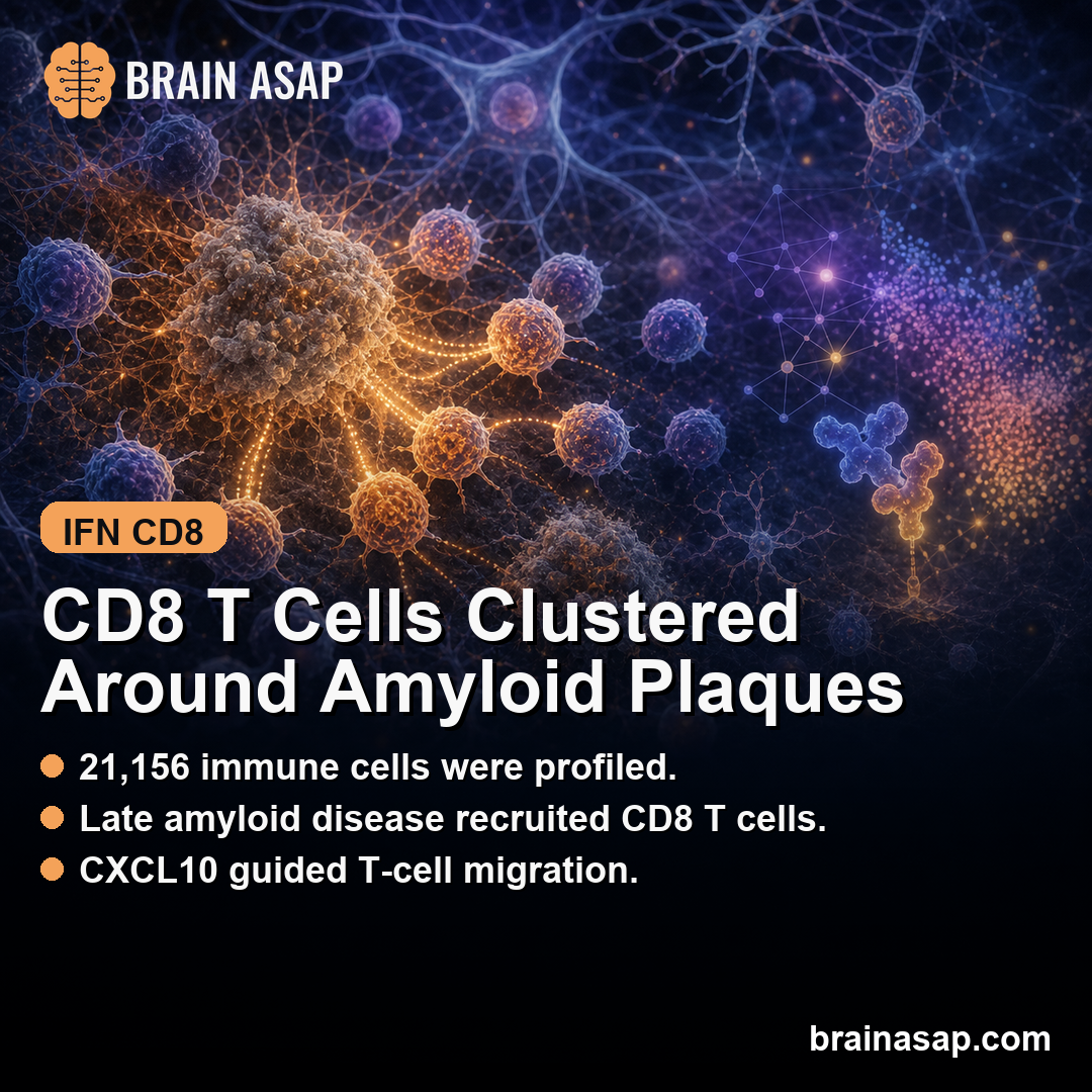
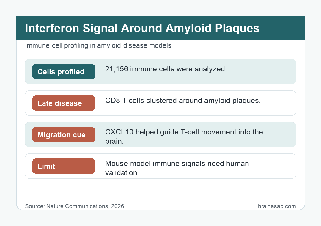
- 21,156 immune cells profiled: The team used single-cell RNA and VDJ sequencing on CD45-positive brain immune cells from APP23 amyloid mice and wild-type littermates.
- Late amyloid disease shifted the immune mix: Early APP23 disease at 12-14 months was more microglia-centered, while 20-24 month disease showed more T-cell representation, especially CD8 T cells.
- T cells clustered around plaques: Histology showed T cells rising with plaque burden, concentrating near amyloid beta plaques, and showing stronger association with parenchymal plaques than vascular amyloid deposits.
- ISG CD8 T cells produced CXCL10: A plaque-associated CD8 T-cell subset expressed interferon-stimulated genes and the chemokine CXCL10, which can attract CXCR3-positive T cells.
- Migration assays supported the mechanism: CXCL10 and ISG-induced CD8 T cells drove T-cell migration in vitro, while CXCR3 knockout or blockade reduced that migration.
Source: Nature Communications (2026) | Michel et al.
Alzheimer’s disease inflammation is usually introduced through microglia, the brain-resident immune cells that surround amyloid plaques and change state as pathology builds.
This study adds a second layer. In an amyloid mouse model and in human postmortem amyloid tissue, the immune neighborhood near plaques also showed signs of CD8 T-cell recruitment, interferon pathway activity, and T-cell activation.
APP23 Amyloid Mice Showed a Late Shift Toward CD8 T Cells
Researchers used APP23 transgenic mice, which carry a human amyloid precursor protein mutation and develop progressive amyloid beta pathology. That model fits this question because it lets researchers study amyloid-driven inflammation without adding tau tangles as a second major pathology.
The design separated amyloid disease into an earlier 12-14 month stage and a later 20-24 month stage. CD45-positive immune cells were isolated from brain tissue, then profiled with single-cell RNA sequencing and T-cell receptor sequencing.
Across 21,156 immune cells, the immune landscape changed with age and amyloid burden.
Early disease had a stronger microglial signature, including disease-associated microglia and interferon-stimulated microglia.
Late disease showed greater lymphocyte activity, with CD8 T cells becoming a more visible part of the plaque response.
The research suggests a sequence: early amyloid inflammation was mostly microglia-centered, while late amyloid inflammation included a more organized CD8 T-cell component.
- Early stage: Microglia dominated the amyloid-linked immune profile.
- Late stage: CD8 T cells became more visible around plaques.
- Migration route: CXCL10-CXCR3 signaling supplied a tested movement mechanism for the T-cell pattern.
CD8 T Cells Concentrated Near Amyloid Beta Plaques
To test whether the T cells were randomly present or actually tied to plaques, the team stained for amyloid beta, CD3-positive T cells, and CD31-positive blood vessels. That allowed them to distinguish parenchymal plaques from vascular amyloid deposits.
T-cell abundance increased with age and plaque burden, peaking around the later amyloid stage. More importantly, the cells were not evenly scattered through the tissue.
They clustered near amyloid beta plaques and were largely absent in plaque-poor regions. The plaque association was stronger for parenchymal amyloid plaques than for vascular amyloid deposits.
Vascular deposits should, in principle, be easier for blood-derived immune cells to access.
The stronger parenchymal association suggests that the local plaque neighborhood, not just vessel access, helped shape T-cell recruitment.
Practical interpretation: the adaptive immune response looked targeted to amyloid-rich tissue rather than being a nonspecific background increase in brain T cells.
Interferon-Stimulated CD8 T Cells Produced CXCL10
The study then narrowed the biology from “more T cells” to a specific inflammatory state.
A subset of CD8 T cells expressed interferon-stimulated genes, often abbreviated as ISGs.
These genes are part of the type I interferon response, a defense program that can be protective during viral infection but damaging when chronically activated in neurodegenerative tissue.
Earlier Alzheimer work has already linked type I interferon activity in microglia to synapse loss and neuroinflammation.
The APP23 results extend that immune pathway to CD8 T cells.
In the APP23 data, ISG microglia were more prominent earlier, while ISG CD8 T cells became more prominent in late amyloid disease.
One molecule made the T-cell result more mechanistic: CXCL10.
CXCL10 is a chemokine, meaning it helps direct immune-cell movement.
The matching receptor, CXCR3, was present on T cells that could be recruited into the inflammatory site.
Mechanism: plaque-associated ISG CD8 T cells appear to release CXCL10, while nearby CXCR3-positive T cells respond to that chemokine gradient and move toward the amyloid-rich tissue.
In the proposed model, ISG CD8 T cells near plaques produce CXCL10, and that CXCL10 helps pull in more CXCR3-positive T cells. The system can therefore amplify itself locally as amyloid disease progresses.
CXCL10-CXCR3 Assays Linked the Spatial Pattern to Cell Movement
Spatial transcriptomics can show where a transcript pattern sits, but it cannot prove that the pattern moves cells. The study added in vitro migration experiments to close that gap.
When recombinant CXCL10 was placed in the receiving chamber of a transwell assay, control CD8 T cells migrated toward it. CXCR3 knockout cells migrated much less, and CXCR3-blocking antibody also reduced chemotaxis.
Researchers then activated CD8 T cells into an interferon-stimulated state using a STING agonist. Those ISG-induced CD8 T cells acted as a strong chemotactic source for control T cells, while migration of CXCR3-deficient T cells was reduced.
The combination is stronger than a marker list because it connects three levels of evidence:
- Single-cell evidence: ISG CD8 T cells and CXCL10 expression were identifiable in late amyloid disease.
- Spatial evidence: CXCL10 and T-cell activation markers were enriched near amyloid plaques.
- Functional evidence: CXCL10-CXCR3 chemotaxis helped drive T-cell movement in migration assays.
CXCL10-CXCR3 is the clearest tested recruitment route in this study, even though other chemokine pathways may also contribute.
Human Alzheimer Tissue Supported the Plaque-Neighborhood Signal
Mouse amyloid models can answer only part of the Alzheimer’s disease question. Researchers therefore tested whether the plaque-neighborhood interferon pattern also appeared in human tissue.
They used postmortem Alzheimer/amyloid tissue cores and targeted spatial transcriptomics. Plaques were segmented into inside, periphery, and outside regions, then transcript density was compared across those zones.
Several interferon-stimulated genes, including IFIT1, IFIT2, and IRF7 , were enriched inside or near plaques. CXCL10 transcripts also concentrated near plaques.
T-cell activation and exhaustion-linked markers, including NKG7, GZMK, and PDCD1, were higher in plaque neighborhoods. That human validation is important, but it should be read carefully.
It supports the idea that amyloid-rich human tissue can carry a local interferon/T-cell activation signature.
It does not prove that these T cells cause cognitive decline in people, nor does it show that blocking the pathway would help patients.
Late Amyloid Plaques Added a T-Cell Neuroinflammation Layer
Alzheimer neuroinflammation is often treated as one process, but the APP23 data separated early and late plaque biology. Early amyloid disease was more microglia-centered, while later amyloid disease included CD8 T cells that clustered around plaques, expanded, activated, and moved toward exhaustion.
That stage difference could help explain why T-cell depletion studies have produced conflicting results. Removing T cells before a full amyloid immune response develops may not have the same effect as targeting a late, plaque-associated, activated CD8 T-cell state.
The therapeutic implication is not “block all T cells in Alzheimer’s.” A better reading is narrower: type I interferon activity and the CXCL10-CXCR3 axis may be stage-dependent targets worth testing in carefully chosen disease windows.
Takeaway: a treatment aimed at plaque-linked T-cell recruitment would probably need to match the stage of amyloid inflammation, not treat early and late disease as the same immune state.
The main limitation is the model.
APP23 mice model amyloidosis but do not reproduce the full Alzheimer’s disease process, especially tau pathology.
The human tissue analysis shows overlap near plaques, but postmortem spatial data cannot establish causality or treatment response.
Still, the study gives a clearer map of amyloid inflammation than a microglia-only frame.
As plaques persist, the immune neighborhood may recruit a second wave: interferon-stimulated CD8 T cells that help pull more T cells into the lesion.
For Alzheimer’s research, the adaptive immune system becomes a time-dependent part of the plaque response, not just a bystander.
Citation: DOI: 10.1038/s41467-026-72262-6. Michel et al. Type I interferon drives T cell responses to amyloid beta in the central nervous system. Nature Communications. 2026;17:3737.
Study Design: APP23 transgenic amyloid mouse model, wild-type littermates, single-cell RNA/VDJ sequencing, histology, targeted spatial transcriptomics, transwell migration assays, and human postmortem Alzheimer/amyloid tissue cores.
Sample/Model: 21,156 profiled immune cells plus APP23 mouse tissue and postmortem human Alzheimer/amyloid tissue cores.
Key Statistic: Late amyloid disease recruited plaque-associated ISG CD8 T cells that expressed CXCL10 and supported CXCR3-dependent T-cell migration in vitro.
Caveat: APP23 mice model amyloidosis rather than the full Alzheimer disease process, and postmortem human spatial data cannot prove treatment response.