TL;DR: A 2026 Nature Methods paper described a self-localized ultrafast pencil beam that made volumetric multiphoton imaging faster and more stable, including 1-minute 3D scans of transferrin uptake in a live human blood-brain barrier model.
Key Findings
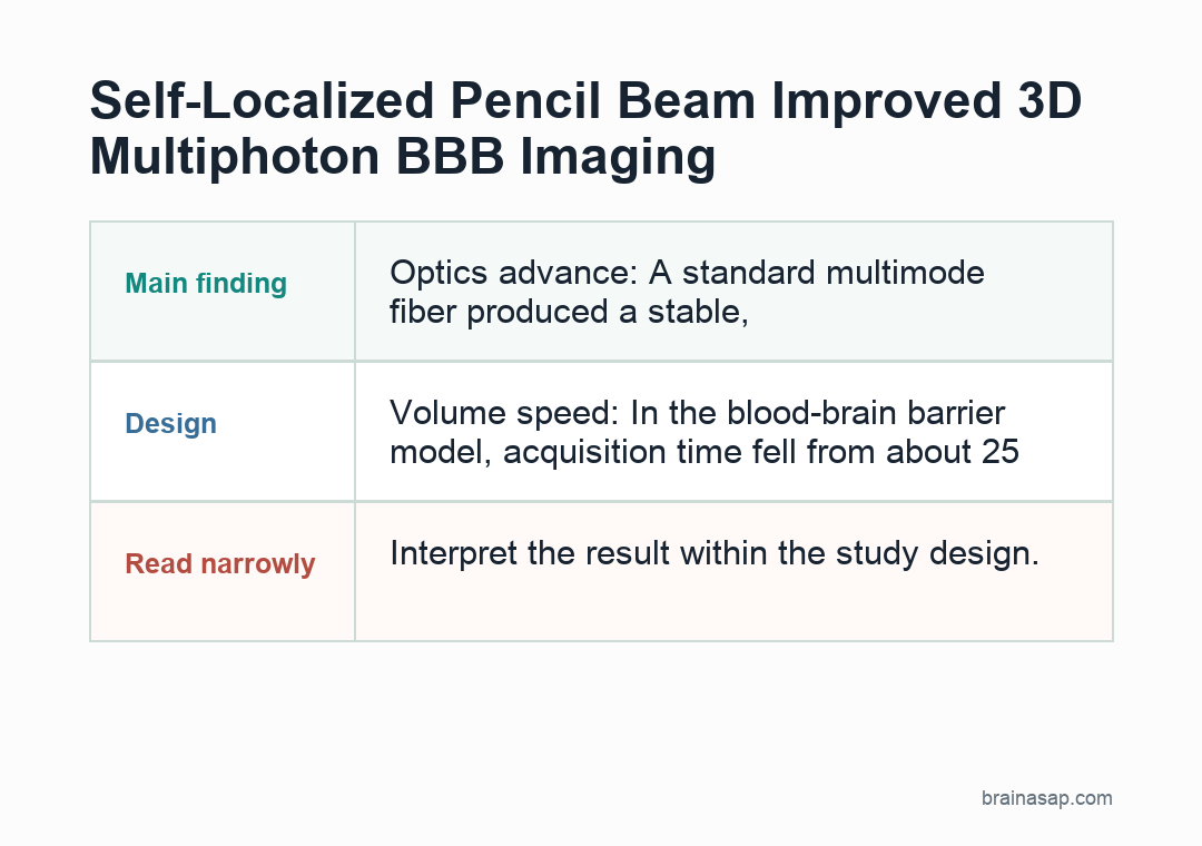
- Optics advance: A standard multimode fiber produced a stable, sidelobe-suppressed Bessel-like pencil beam near critical power.
- Microscopy fit: The beam could be integrated into an existing multiphoton point-scanning microscope.
- Volume speed: In the blood-brain barrier model, acquisition time fell from about 25 minutes to 1 minute for the same 3D volume.
- Biology readout: The system tracked transferrin uptake with NAD(P)H/FAD metabolic phenotyping across 50 minutes.
- Caution: This is mainly a microscopy-methods paper, not evidence that transferrin delivery has been optimized therapeutically.
Source: Nature Methods (2026) | Cao et al.
Volumetric brain and tissue imaging often runs into a speed-versus-resolution problem. A tightly focused Gaussian beam gives sharp detail, but it scans a small focal plane.
Extended-focus beams can cover more depth, but conventional Bessel beams bring sidelobes that add background and blur fine structure.
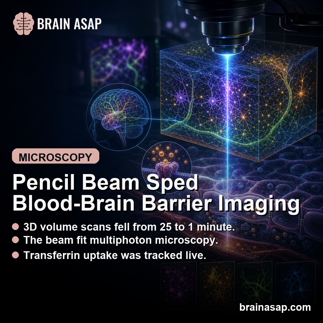
Cao and the research team tested another route: let a standard multimode fiber reorganize high-power ultrafast light into a stable, narrow, extended beam. The study calls it a self-localized ultrafast pencil beam.
In practice, the method aimed to keep the extended depth of a Bessel-like beam while reducing the sidelobe penalty that complicates deep volumetric imaging.
The biological demonstration focused on intact mouse enteric nervous tissue and a live human blood-brain barrier model.
The blood-brain barrier experiment is the easiest place to see why the method could be valuable: the system scanned a 3D volume every minute while tracking how transferrin moved through different cell types.
A Multimode Fiber Generated a Self-Localized Ultrafast Pencil Beam
The optical setup used a step-index multimode fiber, a relatively standard piece of hardware rather than a custom high-complexity beam-shaping system.
By launching an overfilled on-axis Gaussian beam into the fiber near critical power, the researchers observed a nonlinear localized state.
Self-focusing in fibers is often treated as a problem because high peak power can randomize the beam or damage the system.
Here, the fiber’s nonlinear behavior produced a more organized output.
The beam had a Bessel-like profile with suppressed sidelobes, which is exactly the combination needed for faster volumetric point-scanning microscopy.
The optical tests reported improved stability and noise characteristics, including roughly 11 dB noise suppression.
The beam also stayed robust across many fiber-bend configurations, which is important for a method that would otherwise be too fragile for routine microscopy use.
The setup also matters operationally.
The beam was generated by launching light into a standard multimode fiber, then adding it to an existing multiphoton point-scanning microscope.
That keeps the method closer to a retrofit than a completely new microscope platform.
That retrofit angle is part of the appeal.
Advanced microscopy methods often work beautifully in one lab but require too much specialized alignment, custom optics, or active wavefront control for broad use.
A stable beam from a standard fiber lowers that barrier if other labs can reproduce the conditions.
The technical claim depends on three checks:
- Beam shape: extended focus with suppressed sidelobes.
- Stability: robust output across multiple fiber-bend configurations.
- Integration: compatibility with an existing multiphoton point-scanning microscope.
Extended Focus Preserved Resolution Across a 50-Micron Imaging Volume
The key microscopy advantage was axial extension. In a high-numerical-aperture setup, the pencil beam extended the helpful focus along the depth axis while preserving lateral detail well enough for subcellular imaging.
In the blood-brain barrier model, the study reports imaging a 2 mm x 2 mm x 50 micron volume with subcellular spatial resolution.
That is a large field for repeated 3D imaging when the goal is to follow living-cell transport dynamics rather than capture a single endpoint.
The speed comparison was direct.
Conventional sequential sectioning required about 25 minutes for the same volume, while the self-localized pencil-beam approach acquired it in about 1 minute.
That is why the method can shift a slow 3D snapshot into a time-resolved 3D movie.
That time difference is not just a convenience.
Transport events in living barriers can change over minutes, so a 25-minute scan can blur the timing of uptake across cells.
A 1-minute volume makes it easier to compare when endothelial cells, pericytes, and astrocytes begin accumulating a molecule.
The measurement also paired structural imaging with metabolic readouts.
NAD(P)H and FAD autofluorescence are commonly used to estimate cellular redox state, so the setup could follow transport and metabolism in the same live model rather than treating them as separate experiments.
Mouse Enteric Nervous System Imaging Improved at Greater Depth
The first biological benchmark used intact mouse intestinal tissue with tdTomato-labeled enteric neurons. This is a good stress test because tissue scattering and aberration make deep imaging harder, especially for beams with strong sidelobes.
The pencil beam kept lateral resolution comparable to a Gaussian focus while extending axial reach by roughly an order of magnitude.
At greater depths, the self-localized output produced more fluorescence than conventional Bessel-like or speckled outputs.
Those comparisons support the central microscopy claim: the beam was not just longer, it was more practical for tissue imaging.
Reduced sidelobes mean less out-of-focus excitation, and better aberration resilience means the same setup can keep working when tissue distorts the beam.
Live Blood-Brain Barrier Imaging Tracked Transferrin Uptake Every Minute
The blood-brain barrier experiment used a live human model containing endothelial cells, pericytes, and astrocytes.
The researchers monitored fluorescent transferrin uptake while also measuring NAD(P)H and FAD readouts linked to cellular metabolic state.
The time course showed cell-type differences:
- Endothelial cells showed the dominant transferrin uptake and reached a plateau around 45 minutes.
- Pericytes had lower uptake, less than 30% of the endothelial signal.
- Astrocytes showed generally low uptake, with localized hotspots in some areas.
A competitive inhibition experiment strengthened the transport interpretation. Adding unlabeled transferrin after 20 minutes suppressed endothelial accumulation, with about a 5-fold reduction in fluorescence compared with the uninhibited condition.
That inhibition step helps separate receptor-linked uptake from nonspecific fluorescence accumulation. If unlabeled transferrin competes away much of the labeled transferrin pattern, the experiment better supports a transferrin-specific transport process.
The Microscopy Claim Still Needs Biology-Specific Validation
The strongest claim here is technical: the beam enabled faster, more stable volumetric multiphoton imaging.
The biological examples show what the tool can measure, but they do not prove a new blood-brain barrier therapy or a new transport mechanism by themselves.
Several limits are worth keeping in view. The blood-brain barrier system was a model, not a living human brain.
Fluorescent transferrin uptake is a measurable transport process, but therapeutic delivery would require additional drug-cargo, dose, safety, and disease-context testing.
The method also needs broader adoption to show how easily other labs can reproduce the optical state and imaging performance.
Even with those limits, the study is valuable because it links an optical physics advance to a concrete biological measurement.
A 25-fold speed increase can change what scientists are able to observe.
Slow 3D imaging misses fast transport dynamics; minute-resolved 3D imaging can separate cell types, timing, and metabolic state in the same living system.
The near-term use is likely methodological. Labs studying neurovascular transport, organoids, intestinal neural tissue, or other living 3D systems may be able to ask questions that require repeated volumetric scans rather than isolated endpoints.
The strongest next test is reproducibility.
If the pencil-beam state can be generated reliably across microscopes, objectives, fibers, and tissue types, then the study becomes more than a one-system demonstration.
It becomes a practical way to make fast volumetric multiphoton imaging less tradeoff-heavy.
Citation: DOI: 10.1038/s41592-026-03067-0. Cao et al. Self-localized ultrafast pencil beam for volumetric multiphoton imaging. Nature Methods. 2026.
Study Design: Optical-methods study with beam characterization, multiphoton microscope integration, mouse enteric nervous system imaging, and live human blood-brain barrier model imaging.
Sample Size: Not a human participant study; experiments used optical benchmarks, mouse intestinal tissue imaging, and live cell-model imaging.
Key Statistic: Blood-brain barrier 3D volume acquisition was reduced from about 25 minutes to about 1 minute.
Caveat: Microscopy-methods evidence using optical benchmarks, mouse tissue, and a live BBB model; it is not a therapeutic delivery study.


