TL;DR: A 2026 mouse study in Communications Biology found that activating parvalbumin (PV) interneurons, inhibitory nerve cells in the orbitofrontal cortex, reduced default mode network connectivity and decreased normal social approach and sniffing behavior.
Key Findings
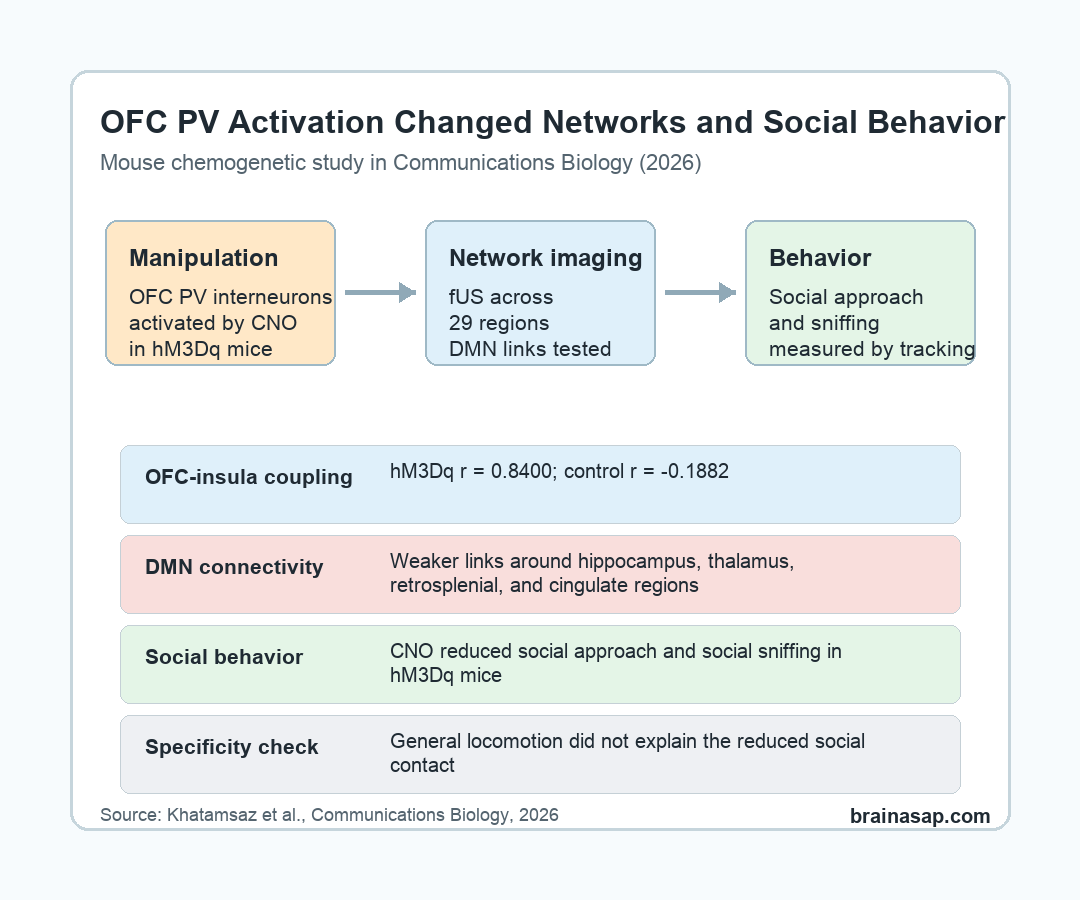
- PV interneurons were targeted: Researchers used PV-Cre mice and a chemogenetic DREADD receptor to activate inhibitory PV interneurons in the orbitofrontal cortex (OFC).
- Functional ultrasound measured networks: Brain-wide cerebral blood volume and connectivity were measured across 29 regions of interest after clozapine-N-oxide administration.
- OFC-insula coupling increased: OFC-insula correlation was strong in the hM3Dq group, r = 0.8400, while the control group correlation was not significant, r = -0.1882.
- Default mode connectivity fell: Activation of OFC PV interneurons reduced connectivity among default mode network regions, especially links involving dorsal hippocampus, subiculum, thalamus, retrosplenial cortex, and cingulate cortex.
- Social behavior decreased without broad motor slowing: CNO reduced social approach and social sniffing in the hM3Dq mice during the dark phase, while general movement did not differ meaningfully between groups.
Source: Communications Biology (2026) | Khatamsaz et al.
Default mode network (DMN) is a set of connected brain regions often studied during rest, self-related processing, and social cognition. Human studies have linked DMN disruption with social impairment in schizophrenia, Alzheimer disease, and other neuropsychiatric conditions.
This mouse study tested a narrower mechanistic question: can changing inhibitory-cell activity in one frontal region push the wider DMN and social behavior in the same direction?
Researchers Activated Orbitofrontal PV Interneurons in Mice
The target was the orbitofrontal cortex (OFC), a frontal brain region involved in decision-making, behavioral regulation, and social-cognitive processing. Researchers focused on PV interneurons, a class of fast-spiking inhibitory cells that can strongly shape local cortical output.
To control those cells, the team used PV-Cre mice carrying an excitatory hM3Dq DREADD receptor. DREADD means designer receptor exclusively activated by designer drug.
In this experiment, clozapine-N-oxide (CNO) was used to activate the engineered receptor.
- Manipulated group: hM3Dq mice had OFC PV interneurons that could be activated by CNO.
- Control group: Control mice received the same general experimental handling without the same active receptor effect.
- Verification step: Researchers used c-Fos staining, a marker of recent neuronal activity, to confirm that the targeted PV interneurons were activated.
The study was preclinical. It does not show that a treatment changes human social behavior.
The experiment tested whether one circuit manipulation can reproduce part of the network and behavior pattern suspected from clinical research.
Functional Ultrasound Tracked 29 Brain Regions After CNO
Researchers used functional ultrasound (fUS), an imaging method that tracks cerebral blood volume as a proxy for local brain activity. The fUS experiment included 13 hM3Dq mice and 11 control mice.
After CNO administration, cerebral blood volume increased selectively in several regions in the hM3Dq group:
- Prelimbic cortex: A medial frontal region involved in behavioral control.
- Orbitofrontal cortex: The manipulated frontal target region.
- Motor cortices: Primary and secondary motor cortex also showed increased blood-volume activity.
The OFC-insula relationship stood out. OFC and insula activity were not meaningfully correlated in controls, with r = -0.1882 and p = 0.5794. In the hM3Dq group, the correlation was r = 0.8400, with p < 0.0001.
Important detail: Increased blood-volume activity in some regions did not mean the whole social network became more connected. The connectivity analysis asked how regions moved together over time.
Default Mode Network Links Were Weaker After PV Activation
The network analysis found lower functional connectivity among DMN-related regions. Researchers calculated correlations across 29 regions of interest and focused on links among OFC, hippocampal, thalamic, retrosplenial, cingulate, and related regions.
After OFC PV activation, connectivity decreased especially around the dorsal hippocampus. Reported affected links included dorsal hippocampus connections with subiculum, somatosensory area, and thalamus, along with additional subiculum-retrosplenial and anterior-cingulate-thalamus links.
- Network direction: The manipulation reduced DMN functional connectivity rather than simply boosting all connected regions together.
- Clinical bridge: Human clinical work has associated DMN dysconnectivity with social impairment.
- Mechanistic limit: The experiment supports a circuit link in mice, not a direct diagnostic marker for people.
The paper also noted a possible pharmacology complication. CNO can be converted to clozapine, and clozapine itself can affect brain-wide connectivity. That caveat is important for interpreting the control-group and drug-route effects.
Social Approach and Social Sniffing Decreased in the hM3Dq Mice
To test behavior, researchers placed familiar groups of 4 mice into a social arena and used combined RFID and video tracking. After 3 days of habituation, mice were tested on vehicle and CNO days.
The behavioral readout focused on 2 social behaviors: social approach and social sniffing. CNO significantly reduced both behaviors in the hM3Dq group during the first 6 hours of the dark phase and across the full 12-hour dark phase.
Controls did not show the same CNO-versus-vehicle difference. The group-specific result ties the behavioral change to the targeted OFC PV interneuron manipulation rather than to the injection schedule alone.
- Social approach fell: hM3Dq mice spent less time approaching other mice after CNO.
- Social sniffing fell: hM3Dq mice also spent less time in a direct social-investigation behavior.
- General locomotion held: Movement data did not indicate a broad motor deficit that would explain less social contact.
Novel Object Testing Suggested a Cognitive Effect Too
Researchers also used a novel object recognition test to check whether the mice were generally inactive or unable to explore. The hM3Dq mice showed object exploration time similar to controls, which argued against a simple activity-level explanation.
Still, only control mice explored the novel object significantly more than the familiar object. The hM3Dq mice did not show the same novelty preference, suggesting that OFC PV activation may have affected object-memory-related behavior as well as social behavior.
That finding fits the broader network interpretation. A frontal inhibitory-cell manipulation did not only change a local signal; it altered large-scale connectivity and behaviors that depend on distributed circuits.
OFC PV Activation Links Mouse DMN Changes With Social Behavior
The study connects 3 levels of evidence in one experiment: targeted OFC inhibitory-cell activation, DMN connectivity changes, and reduced social behavior.
The result should stay bounded. Mouse social behavior in a controlled arena is not the same as human social withdrawal, schizophrenia, Alzheimer disease, or autism-related social difficulty.
- Species limit: The experiment used mice, so human clinical implications require separate testing.
- Drug-route limit: CNO-to-clozapine conversion can complicate interpretation of connectivity effects.
- Behavior limit: Social approach and sniffing are measurable mouse behaviors, not full analogs of human social relationships.
- Strength: The same manipulation linked OFC inhibitory cells, DMN connectivity, and social behavior in one controlled design.
For now, the mechanistic takeaway is specific: activating OFC PV interneurons was enough to weaken DMN connectivity and reduce normal social interaction behaviors in mice.
Citation: DOI: 10.1038/s42003-026-10060-y. Khatamsaz et al. Orbitofrontal PV interneurons modulate social interaction via default mode network dynamics. Communications Biology. 2026;9:573.
Study Design: Mouse chemogenetic experiment combining OFC PV interneuron activation, functional ultrasound connectivity imaging, and automated social-behavior tracking.
Sample/Model: PV-Cre mice with hM3Dq DREADD manipulation; fUS analysis included 13 hM3Dq mice and 11 controls.
Key Statistic: OFC-insula correlation was r = 0.8400 in the hM3Dq group versus r = -0.1882 in controls, and social approach and sniffing decreased after CNO in hM3Dq mice.
Caveat: The study is a mouse circuit experiment, and CNO conversion to clozapine can complicate interpretation of brain-wide connectivity effects.


