TL;DR: A 2026 Current Biology study found that bacterial polysaccharides, including peptidoglycan from Gram-positive bacteria, activated the C. elegans NSM enteric sensory neuron and shifted feeding and movement behavior through acid-sensing ion channels.
Key Findings
- NSM sensed ingested bacteria: the enteric sensory neuron NSM, which projects into the C. elegans pharyngeal lumen, responded when worms ingested diverse bacteria.
- Polysaccharides were sufficient: bacterial polysaccharides activated NSM after researchers separated bacterial macromolecules and tested candidate fractions.
- Peptidoglycan was one specific food cue: peptidoglycan from Gram-positive bacteria activated NSM, giving the circuit a concrete molecular input rather than a vague microbiome association.
- DEL-3 and DEL-7 were required: NSM responses to polysaccharides depended on acid-sensing ion channels that localize to the neuron’s sensory dendrite.
- Prodigiosin blocked the food response: the pathogenic Serratia marcescens pigment prodigiosin prevented NSM activation by nutritive bacterial cues.
Source: Current Biology (2026) | Estrem et al.
C. elegans Turned Gut-Brain Signaling Into a Testable Circuit
Microbiome research often starts with associations: one bacterial community tracks with one disease state or behavior. This study asked a narrower question: which bacterial molecules can a nervous system detect directly?
Researchers used Caenorhabditis elegans, a transparent nematode that eats bacteria. The model is useful because the animal has a compact nervous system, a bacterial diet, and feeding behaviors that can be measured when a sensory neuron turns on.
The target neuron was NSM, an enteric sensory neuron that innervates the pharyngeal lumen, the part of the worm’s feeding apparatus where bacteria pass during ingestion. When NSM is activated, it releases serotonin and changes feeding-related behavior.
Polysaccharides Activated the NSM Feeding Neuron
The team first tested whether different bacteria activated NSM. They then broke bacteria into major molecular classes to determine which class the neuron was detecting.
The result pointed to bacterial polysaccharides, sugar-based molecules that can sit on bacterial surfaces. DNA, lipids, proteins, and simple sugars were not the main explanation in the same screening logic.
Researchers narrowed one specific bacterial cue further:
- Broad bacterial cue: polysaccharides from bacteria were sufficient to activate NSM.
- Gram-positive cue: peptidoglycan, a structural polymer in bacterial cell walls, activated NSM as a defined component.
- Behavioral match: ingestion of these bacterial polysaccharides increased feeding and reduced locomotion, matching known NSM-driven behaviors.
The behavioral match is important. The response was not just visible in a calcium trace or cell assay; it matched the animal’s feeding program.
DEL-3 and DEL-7 Carried the Sensory Channel Requirement
The neural detection step depended on DEL-3 and DEL-7, two acid-sensing ion channels. These channels localized to the NSM sensory dendrite in the pharyngeal lumen, where ingested bacterial material would be encountered.
When the relevant channel function was removed, NSM responses to bacterial polysaccharides were lost. That places the bacterial cue, sensory channel, neuron, and behavior in the same causal chain.
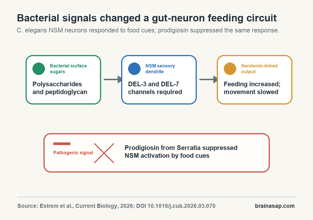
The study’s mechanism can be read as four linked steps:
- Bacterial ingestion: the worm takes nutritive bacteria into the pharyngeal lumen.
- Molecular detection: bacterial polysaccharides, including peptidoglycan, reach the NSM sensory ending.
- Channel-dependent activation: DEL-3 and DEL-7 are needed for the NSM response.
- Behavioral output: NSM serotonin signaling promotes feeding and slows locomotion.
Prodigiosin From Serratia Suppressed the Food Response
The pathogenic-bacteria experiment added a second side to the circuit. Researchers tested Serratia marcescens, a bacterium that can infect C. elegans and can produce a red pigment called prodigiosin.
Non-pigmented Serratia still activated NSM and was ingested. Prodigiosin-containing bacteria did not trigger the same NSM response, and adding prodigiosin to otherwise nutritive bacteria suppressed the usual activation.
The worm was not only detecting that bacteria were present. The same neuron appears to integrate competing bacterial cues: food-associated polysaccharides and danger-associated prodigiosin.
The Mechanism Does Not Prove a Human Microbiome Treatment
The finding does not show that bacterial sugars control human mood or that one supplement can tune the gut-brain axis. C. elegans is a simplified model with a specialized bacterial diet.
The value is mechanistic. The study identified a concrete route by which bacterial molecules can be sensed by a neuron and translated into behavior.
That is still a meaningful step for the field. It gives later mammalian studies a specific set of candidate cues and receptors to test instead of treating gut bacteria as an undifferentiated exposure.
Three boundaries keep the interpretation proportional:
- Species boundary: C. elegans has a much simpler nervous system than mammals, and its feeding circuit is specialized for bacterial food.
- Behavior boundary: the measured outputs were worm feeding and locomotion, not anxiety, depression, cognition, or Parkinson’s disease.
- Translation boundary: related molecular players exist across species, but mammalian gut-brain signaling has additional immune, endocrine, vagal, and microbial layers.
Specific Bacterial Cues Give Gut-Brain Research a Cleaner Starting Point
The strongest implication is that neurons can detect specific bacterial structures, not only respond secondhand after immune or intestinal cells react. That gives gut-brain research a cleaner molecular starting point.
The practical interpretation should stay modest: this is a foundational neural-circuit study, not a clinical microbiome intervention. It helps explain how bacterial identity could be converted into nervous-system activity in an animal built to read bacteria as food or threat.
The work also shows why gut-brain experiments need molecular specificity. “The microbiome” is too broad to explain behavior by itself; this study points to named bacterial cell-wall molecules, named ion channels, a named neuron, and a defined pathogen pigment.
Citation: DOI: 10.1016/j.cub.2026.03.070. Estrem et al. Identification of bacterial signals that modulate enteric sensory neurons to influence behavior in C. elegans. Current Biology. 2026.
Study Design: C. elegans neural-circuit and behavior study using bacterial-fraction testing, neuron-activation assays, channel-dependence experiments, and feeding/locomotion behavior readouts.
Sample/Model: Caenorhabditis elegans NSM enteric sensory neuron and bacterial feeding/pathogen-exposure paradigms.
Key Statistic: Bacterial polysaccharides were sufficient to activate NSM, DEL-3 and DEL-7 were required for the response, and prodigiosin suppressed activation by nutritive bacterial cues.
Caveat: Worm feeding behavior is a mechanistic model; the study does not test human mood, cognition, or clinical microbiome treatment.