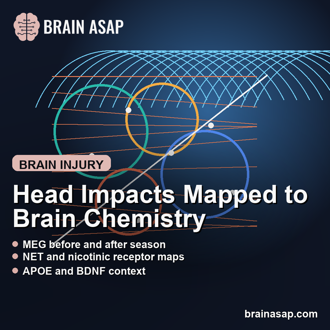
TL;DR: A 2026 preprint in medRxiv found that head-impact-related magnetoencephalography (MEG) changes in high-school football players were strongest in cortical regions matching specific neurotransmitter and gene-expression maps, especially norepinephrine, nicotinic acetylcholine, APOE, and BDNF-related patterns.
Key Findings
- MEG tracked pre-to-post-season brain signaling across 278 timepoints from 91 male high-school football players.
- Concussion-related cortical slowing aligned most strongly with norepinephrine transporter and alpha-4 beta-2 nicotinic acetylcholine receptor maps.
- Cognitive symptom links appeared in the same chemistry-rich regions, with PCSI cognitive scores aligning with NET and alpha-4 beta-2 receptor density.
- Non-concussive head impacts showed a related but narrower pattern, aligning with 5-HT1A serotonin receptor density plus APOE and BDNF expression.
- The source is a preprint, and the concussion subgroup included only 10 players, so the finding provides mechanistic context rather than a clinical test.
Source: The preprint combined magnetoencephalography, helmet-sensor head-impact exposure, symptom scores, neurotransmitter atlases, and Allen Human Brain Atlas gene-expression maps.
MEG Showed Where Football Head Impacts Changed Cortical Activity
Magnetoencephalography (MEG), a brain-imaging method that records fast magnetic fields from neural activity, was used to compare each athlete’s brain activity before and after football seasons. That design let researchers ask where head impacts changed cortical signaling rather than relying on one post-injury scan.
The dataset covered 91 male high-school football players across up to four seasons, producing 278 pre- and post-season timepoints. Ten players had a clinically diagnosed concussion during a measured season.
For players without diagnosed concussion, helmet-mounted sensors estimated cumulative head-impact exposure through a risk-weighted exposure score. That gave the researchers two related comparisons: diagnosed concussion and repeated non-concussive impacts.
- Concussion model: Compared seasons with diagnosed concussion against non-concussion seasons while adjusting for age, time between scans, and body mass index.
- Non-concussive model: Tested whether higher cumulative impact exposure predicted similar pre-to-post-season MEG shifts.
- Symptom model: Linked cognitive symptom scores to the cortical pattern of MEG change in the concussed players.
The main MEG pattern was aperiodic slowing. In plain terms, the researchers treated a steeper aperiodic exponent as a marker of lower cortical excitability, meaning the affected cortex looked less electrically excitable after exposure.
Concussion Slowing Aligned With Norepinephrine and Nicotinic Receptor Maps
The strongest point was not simply that concussion changed brain activity. The important detail was where those changes were strongest.
Concussion-related aperiodic slowing aligned with higher atlas-based density of the norepinephrine transporter (NET) and the alpha-4 beta-2 nicotinic acetylcholine receptor. The correlations were moderate to strong for a brain-map analysis: r = .43 for NET and r = .50 for alpha-4 beta-2 receptor density after false-discovery-rate correction.
Those systems are not random choices. Norepinephrine helps regulate arousal, attention, and wakefulness, while acetylcholine is central to attention and memory. Both have long been discussed in traumatic brain injury biology.
The same concussion-related slowing also aligned with several other neurotransmitter maps, including vesicular acetylcholine transporter, 5-HT1B serotonin receptor, and histamine H3 receptor density. In the opposite direction, slowing was stronger where several dopamine and serotonin markers were lower, including D2 dopamine receptor density.
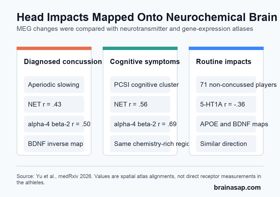
The gene-expression layer added another boundary: regions with higher BDNF, a gene tied to neurotrophic support and plasticity, were relatively less aligned with concussion-related slowing, and APOE expression showed an inverse alignment too.
Cognitive Symptoms Followed the Same Chemistry-Rich Regions
The symptom analysis made the MEG result more clinically relevant, although it remained small. Caregiver-rated cognitive symptoms came from the Post-Concussive Symptom Inventory (PCSI), including problems such as feeling mentally foggy, slowed down, confused, or less able to concentrate.
Among the concussed players, stronger links between aperiodic slowing and cognitive symptoms again aligned with regions rich in NET and alpha-4 beta-2 receptor density. The reported alignments were r = .56 for NET and r = .69 for alpha-4 beta-2 receptor density.
The symptom link connects the imaging change to a clinically relevant map instead of leaving the MEG result as an abstract brain measurement.
- Brain signal: MEG showed lower-excitability-like slowing after concussion.
- Chemical context: The slowing was strongest in regions matching norepinephrine and nicotinic acetylcholine maps.
- Symptom context: Cognitive symptom severity followed a similar regional pattern.
NET or nicotinic receptors were not shown to cause the symptoms. The atlases came from normative adult brain resources, so they show spatial context, not direct neurotransmitter measurements in these teenage athletes.
Routine Head Impacts Shared Part of the Same Molecular Map
Non-concussive impact exposure did not reproduce every concussion finding. It did, however, point in the same broad direction for aperiodic slowing.
In 71 non-concussed players with complete imaging and kinematic data, aperiodic slowing linked to cumulative head-impact exposure aligned with 5-HT1A serotonin receptor density, APOE expression, and BDNF expression. The direction of the APOE and BDNF effects matched the diagnosed-concussion analysis.
Reduced alpha-band rhythm after non-concussive impacts also aligned with several of the same systems seen in concussion-related alpha changes, including alpha-4 beta-2 receptor density, histamine H3 receptor density, mu-opioid receptor density, APOE expression, and CCR5 expression.
- Diagnosed concussion: The clearest aperiodic slowing map centered on norepinephrine and nicotinic acetylcholine systems.
- Non-concussive exposure: The aperiodic map was narrower and included 5-HT1A, APOE, and BDNF.
- Alpha rhythms: Rhythmic alpha changes appeared more tied to inhibitory, arousal, opioid, and inflammatory-map context than to one single transmitter.
Repeated head impacts may affect cortical activity in regions with molecular profiles already relevant to injury biology, even when no concussion is diagnosed.
The Limits Are the Small Concussion Group and Atlas-Based Biology
The most important limitation is sample size. Only 10 players had diagnosed concussion during a measured season.
Repeated pre-to-post-season scans reduce some noise, but they do not remove the uncertainty that comes with a small concussion subgroup.
The sample also included male high-school football players from one league. Results may not generalize to female athletes, other sports, younger children, adults, or athletes with different impact patterns.
The molecular maps were indirect. Neuromaps and Allen Human Brain Atlas data describe normative neurotransmitter and gene-expression topographies, not the players’ own receptor binding, transporter density, or gene expression after injury.
- No direct chemistry assay: MEG was compared with atlas maps rather than receptor-imaging or biochemical measurements from the same athletes.
- Preprint status: The paper had not been peer-reviewed at the time of posting.
- Sensor boundary: Helmet-mounted sensors are useful but less tightly coupled to the skull than newer mouthpiece-based systems.
Head impacts did not produce a uniform brain-wide pattern. The strongest changes appeared in cortical regions whose chemistry and gene-expression profiles already make sense in traumatic brain injury biology.
Citation: DOI: 10.64898/2026.04.09.26350342. Study authors et al. Neurochemical and genetic organization of head impact effects on cortical neurophysiology. medRxiv. 2026.
Study Design: Longitudinal pre-to-post-season MEG analysis with head-impact sensor exposure, symptom scores, neurotransmitter atlases, and gene-expression maps.
Sample Size: 91 male high-school football players, 278 pre/post-season timepoints, 10 diagnosed concussion cases, and 71 non-concussed players with complete imaging and kinematic data.
Key Statistic: Concussion-related aperiodic slowing aligned with NET density (r = .43) and alpha-4 beta-2 nicotinic receptor density (r = .50); cognitive symptom-linked slowing aligned with alpha-4 beta-2 density at r = .69.
Caveat: Preprint findings, small concussion subgroup, all-male football sample, and atlas-based molecular inference mean this is not yet a clinical diagnostic test.