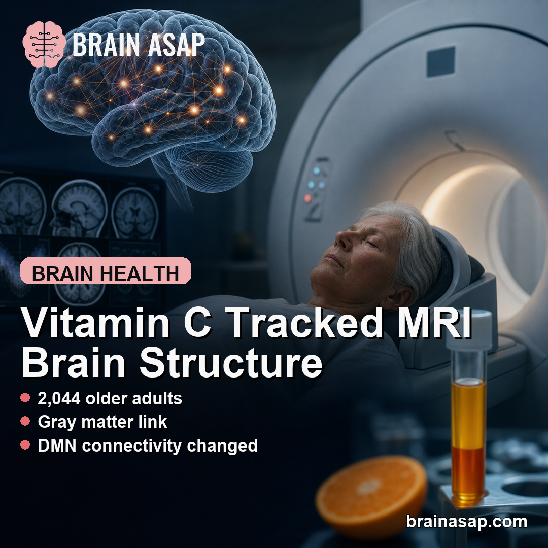
TL;DR: A 2026 cohort study in PLOS One found that higher fasting plasma vitamin C levels were independently associated with larger MRI-measured gray matter and stronger default mode network (DMN) structural connectivity in 2,044 older adults, but the cross-sectional design cannot show that vitamin C caused the brain differences.
Key Findings
- Large MRI cohort: The analysis used 3T brain MRI and fasting blood samples from 2,044 community-dwelling Japanese participants with a median age of 69 years.
- Gray matter link: Plasma vitamin C was independently associated with the gray matter volume/intracranial volume ratio (beta = 0.076; P < 0.001).
- White matter link: Plasma vitamin C was also associated with the white matter volume/intracranial volume ratio (beta = 0.074; P < 0.001).
- DMN connectivity: Vitamin C levels were associated with three default mode network components after correction for multiple comparisons.
- Main caveat: The study measured association at one time point, so it cannot separate vitamin C biology from diet, health status, or other unmeasured factors.
Source: PLOS One (2026) | Nagaya et al.
Plasma vitamin C was linked to several MRI markers of brain structure in a large older-adult cohort. The study does not prove that vitamin C supplementation protects the brain, but it does put a measurable blood nutrient beside MRI-derived brain volumes and structural network measures.
The researchers analyzed 2,044 participants from the Iki-Iki study. Participants had 3T magnetic resonance imaging, fasting plasma vitamin C measurement, Mini-Mental State Examination scoring, and clinical/lifestyle covariates including diabetes, hypertension, hyperlipidemia, smoking, drinking, education, and physical activity.
Higher Plasma Vitamin C Tracked With Larger Gray Matter Volume
The clearest volume result involved the gray matter volume/intracranial volume ratio. This ratio compares gray matter volume with total head size, so the result is less likely to reflect simple differences in skull or brain size.
After adjustment, plasma vitamin C was positively associated with the gray matter ratio (beta = 0.076; standard error, 0.018; P < 0.001). A separate Spearman correlation showed the same direction (rho = 0.196; P < 0.001).
- Sample size: The MRI analysis included 2,044 people after 346 exclusions from the original 2,390 participants.
- Age profile: Median age was 69 years, with an interquartile range of 67 to 73 years.
- Measured blood level: Median plasma vitamin C was 7.38 micrograms per milliliter.
The white matter result pointed in the same direction. Plasma vitamin C was associated with the white matter volume/intracranial volume ratio (beta = 0.074; P < 0.001), although the simple correlation was weaker than for gray matter.
Vitamin C Was Also Associated With Default Mode Network Measures
The study also examined the default mode network (DMN), a set of brain regions often active during internal thought, memory, and self-referential processing. The authors used source-based morphometry, an MRI analysis method that identifies shared gray matter patterns across participants.
Independent component analysis produced 147 gray matter structural networks. Three were interpreted as DMN components: one anterior DMN component, one posterior DMN component involving the posterior cingulate cortex and precuneus, and a second posterior DMN component that also included inferior parietal and lateral temporal cortices.
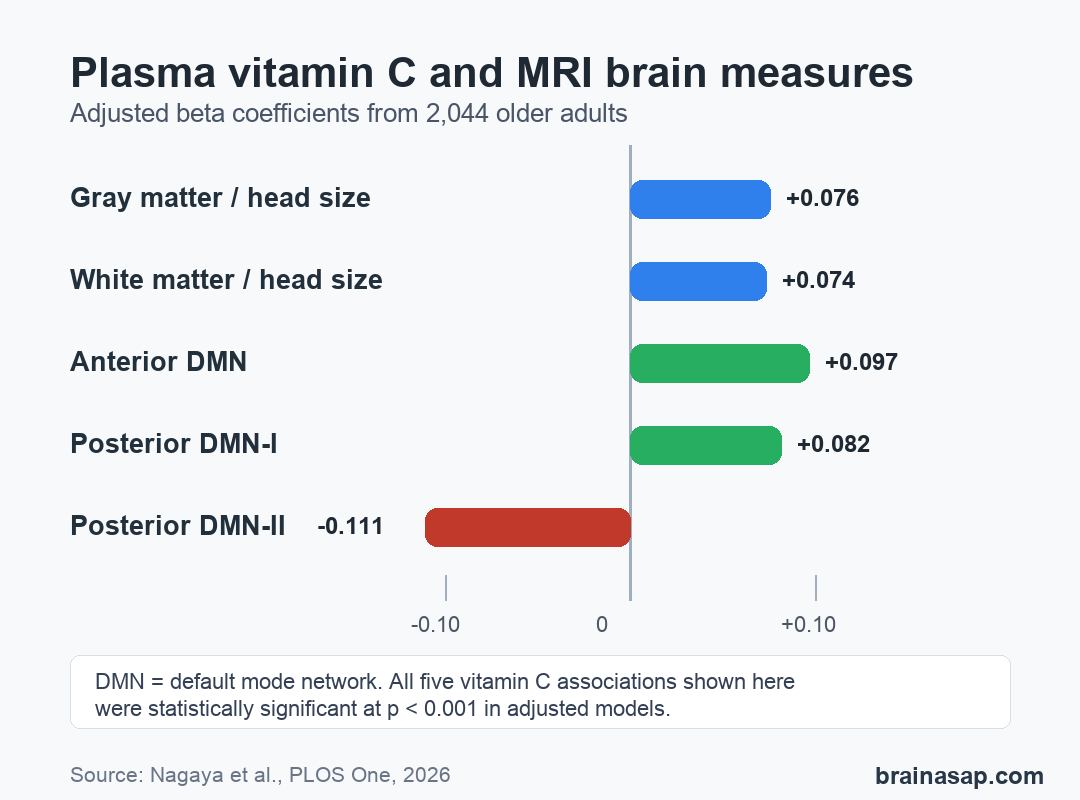
Plasma vitamin C was significantly associated with all three DMN components after false-discovery-rate correction. The adjusted coefficients were 0.097 for anterior DMN, 0.082 for posterior DMN-I, and -0.111 for posterior DMN-II, all with P < 0.001.
- Anterior DMN: Higher vitamin C was associated with a higher component loading coefficient.
- Posterior DMN-I: Higher vitamin C was also associated with a higher loading coefficient in a component including the posterior cingulate cortex and precuneus.
- Posterior DMN-II: The association was negative, which makes the result less straightforward than a simple “more connectivity is better” claim.
That mixed direction matters. The paper supports an MRI association, not a simple consumer-health message that more vitamin C automatically improves every brain network.
The Models Adjusted for Major Health and Lifestyle Factors
The researchers adjusted for several factors that could otherwise blur the vitamin C result. These included age, sex, education, Mini-Mental State Examination score, diabetes, hypertension, hyperlipidemia, smoking history, drinking history, and physical activity.
Some covariates had their own associations. Age was strongly linked to both gray and white matter ratios.
Diabetes was associated with lower gray matter ratio, and smoking history was associated with lower white matter ratio.
- Diabetes: Diabetes had a negative association with the gray matter ratio in the adjusted model.
- Smoking history: Smoking history was associated with the white matter ratio.
- Education and MMSE: The models included education and Mini-Mental State Examination score, a brief cognitive screening test, to reduce confounding by cognitive and social factors.
Adjustment makes the vitamin C association more specific, but it does not remove all confounding. Vitamin C blood levels can reflect broader diet quality, health behaviors, inflammation, illness burden, or access to nutritious food.
This Is Not a Supplement Trial
The safest interpretation is narrow: in this cohort, lower plasma vitamin C went with less favorable MRI brain structure and DMN measures after adjustment. The study does not show whether changing vitamin C intake would change MRI outcomes.
The authors named several limits. The analysis was cross-sectional, used one fasting vitamin C measurement, and came from older Japanese residents with a relatively high education profile.
The models also did not include every possible lifestyle or nutritional factor, such as body mass index or total dietary intake.
- No causality: Brain structure and vitamin C were measured at the same time.
- Single blood draw: One fasting sample may not represent long-term vitamin C status perfectly.
- Population boundary: Results may not generalize directly to younger adults or more diverse populations.
For brain-health research, the paper is useful because it connects a measurable nutrient biomarker with MRI structure in a large cohort. For clinical advice, it is not enough to prescribe vitamin C supplementation as a brain-structure intervention.
Citation: DOI: 10.1371/journal.pone.0348504. Nagaya et al. Plasma vitamin C levels are associated with brain structural networks on MRI: A large cohort study. PLOS One. 2026;21(6):e0348504.
Study Design: Cross-sectional cohort analysis using fasting plasma vitamin C and 3T MRI measures.
Sample Size: 2,044 community-dwelling older adults from the Iki-Iki study.
Key Statistic: Plasma vitamin C was associated with gray matter volume/intracranial volume ratio (beta = 0.076; P < 0.001) and three DMN components after multiple-comparison correction.
Caveat: One-time observational measurement cannot prove that vitamin C caused differences in brain volume or network structure.