The Energy Paradox: Why Depressed Brains Burn Faster
TL;DR: Young adults with depression produce ATP—the cell’s energy currency—at faster rates in both brain and blood, yet still feel more fatigued. New research reveals this is a compensatory mechanism that works at rest but collapses under stress, opening a fresh window into the biology of depression-related fatigue.
Fatigue in major depression is relentless and disabling, yet it remains poorly understood. Now a new study reveals an unexpected culprit: not a shortage of energy production, but cells that must work harder to sustain baseline function. Researchers used cutting-edge brain imaging to measure ATP synthesis directly in young adults with and without depression, uncovering a cellular paradox that reframes how we think about depressive exhaustion.
Key Findings
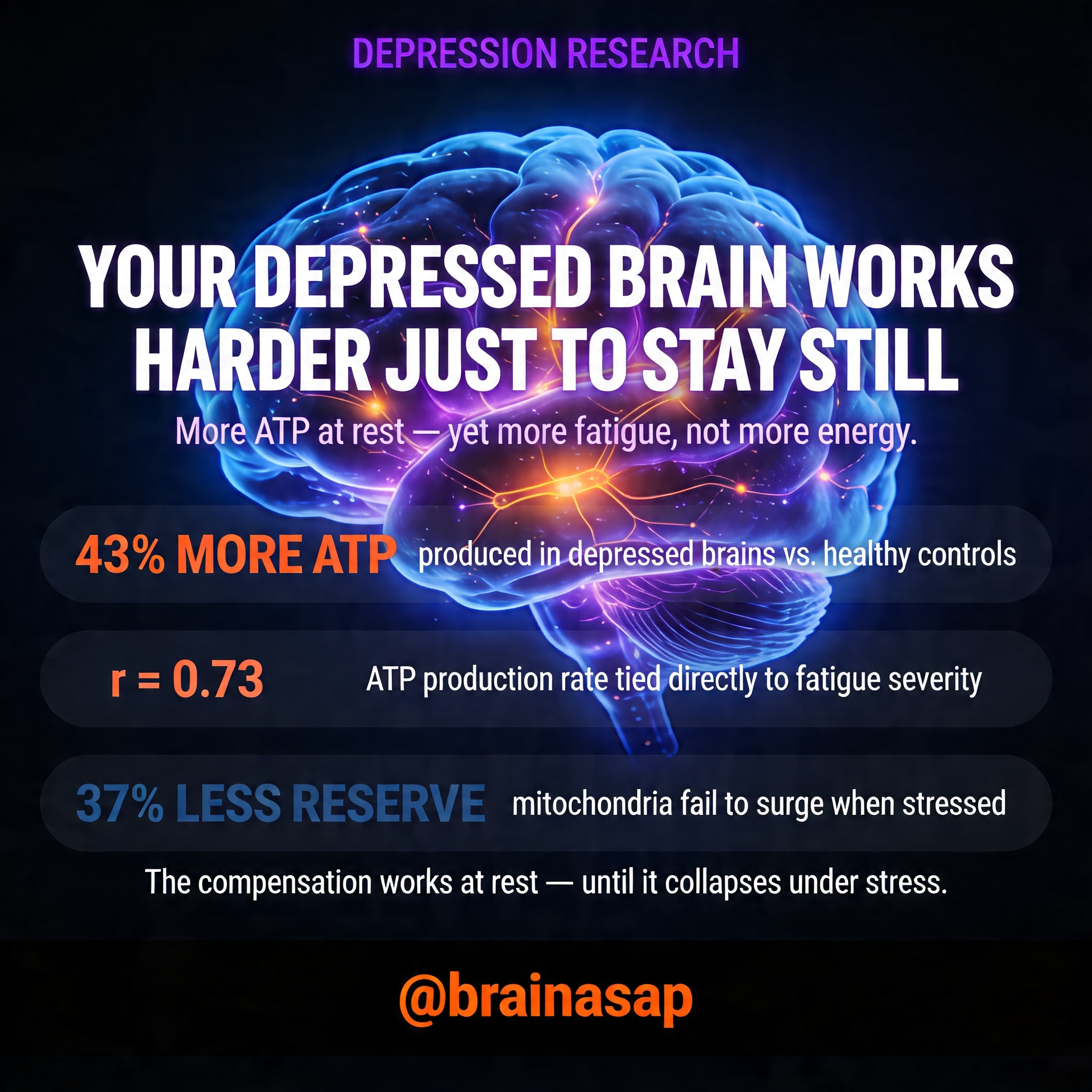
- Higher brain ATP production: The depression group showed 43% higher ATP production rates in the visual cortex (27.5 vs 19.2 μmol/g/min, p=0.004) despite no difference in total ATP concentration.
- Blood ATP mirrors brain: Peripheral blood cells from the depression group had significantly elevated ATP levels (p=0.001) that correlated strongly with fatigue severity (r=0.58, p=0.004).
- Fatigue-energy correlation: The strongest link was between ATP production rate and fatigue scores (r=0.73, p=0.0005)—the harder cells worked to make ATP, the more fatigued participants reported.
- Impaired stress response: When blood cells were stressed with mitochondrial uncouplers, depression participants showed 37% lower spare respiratory capacity, indicating mitochondria unable to ramp up energy production during demand.
- Brain-blood connection: ATP concentrations in blood cells correlated with ATP production rates in the brain (r=0.52, p=0.033), suggesting systemic dysfunction in energy metabolism.
- Sample and context: 18 participants (9 per group, mean age 21.8 years) with usable brain imaging; 24 with blood data; MDD group met criteria for at least moderate depression severity.
Source: Translational Psychiatry (2026) | Cullen et al.
The Brain’s Energy Budget: Why ATP Matters for Depression
The brain consumes an astonishing amount of energy. Though it represents only 2% of body weight, it devours roughly 20% of the body’s glucose and oxygen. This insatiable demand reflects one fundamental need: maintaining ATP—adenosine triphosphate—the molecular currency that powers every neural process from firing synapses to maintaining cell membranes.
For decades, depression research has theorized that fatigue stems from energy metabolism gone wrong. Yet no one had directly measured ATP production rates in the living human brain of depressed individuals. Most prior studies used older imaging techniques that could only measure ATP concentration—like measuring money in a wallet without tracking income or spending. This new research cracked open that black box using a breakthrough technique: phosphorus-31 magnetic resonance spectroscopy with magnetization transfer (31P MRSI-MT) at 7 Tesla.
Compensatory Overdrive: The Cellular Exhaustion Loop
The study recruited 25 young adults—9 with moderate-to-severe depression, 9 healthy controls. Researchers scanned the visual cortex of each participant at ultrahigh field strength and drew blood to analyze mitochondrial function. The results overturned expectations.
Rather than producing less ATP, the depression group produced more ATP—significantly more. Their rate constant for ATP synthesis (kf,ATPase) and cerebral metabolic rate (CMRATPase) were both elevated. At first glance, this seems backwards: if depression correlates with more ATP production, shouldn’t fatigue decrease? The answer lies in understanding the quality of that compensation.
The researchers propose a model: early in depression, cells upregulate ATP production to maintain baseline energy stability in the face of metabolic stress. ATP concentrations remain normal at rest. But this compensation comes at a cost. When cells face increased demand—like during mental exertion or immune stress—they fail. In stress tests using mitochondrial uncouplers (drugs that force cells to work harder), depression participants’ blood cells showed markedly lower oxygen consumption and ATP production capacity than controls.
This is a system that can sustain the moment but not handle a sprint. The fatigue, then, may be adaptive: the brain and body literally cannot afford higher metabolic demands, so fatigue serves as a brake, conserving energy for essential functions.
The Brain-Blood Connection: A Systemic Signature
One of the study’s most striking contributions is demonstrating that ATP dysfunction appears not just in the brain but also in peripheral blood cells. When researchers measured ATP in peripheral blood mononuclear cells (PBMCs), they found higher resting ATP concentrations in the depression group—mirroring the brain findings. More remarkably, blood ATP levels correlated directly with visual cortex ATP production rates, suggesting a systemic bioenergetic signature rather than a localized brain problem.
This opens a practical window. Brain imaging at 7 Tesla is expensive, specialized, and not widely available. But testing blood ATP is quicker, cheaper, and accessible. If blood ATP measures can reliably indicate brain energy metabolism, they could become a screening tool for depression-related fatigue or a biomarker for treatment response.
The strongest evidence for the energy-fatigue link came from correlations: ATP production rate in the brain correlated most strongly with fatigue severity scores (r=0.73). Those making the most ATP reported the highest fatigue. The chemistry speaks clearly: cells forced into metabolic overdrive experience accelerated wear, and the conscious experience is exhaustion.
Why Young Adults? And What Happens Later?
This study focused on young adults (mean age 21.8 years) early in their depression history. That matters. The researchers propose that what we’re seeing is an early compensatory phase—cells upregulating ATP production to maintain function despite metabolic strain. But compensation has limits.
There’s an intriguing parallel to Parkinson’s disease: an earlier study using the same 31P MRSI-MT technique found that early-stage Parkinson’s patients also showed elevated ATP production rates in the visual cortex. In both conditions, young patients show this elevated metabolic machinery trying to keep things running. The implication is sobering: this compensatory mechanism may itself be a risk factor. The cells are working harder, accumulating stress, and eventually—in chronic or recurrent depression, or across decades—may fail entirely.
Studies in older adults (ages 50–69) with depression show the opposite pattern: lower ATP levels in blood cells. This suggests a potential disease progression: early upregulation, then eventual collapse into energy deficit and chronic disability. Longitudinal studies will be essential to test this hypothesis.
Implications for Treatment and Future Research
If ATP bioenergetics is genuinely implicated in depression fatigue, it opens new research avenues. A few prior studies hint at this possibility: one found that beta-ATP levels in the brain predicted treatment response to fluoxetine, while another showed beta-ATP increased in depression patients who responded to thyroid hormone augmentation. No clinical trial yet has used the newer kinetic measures (CMRATPase and kf,ATPase), which this research suggests are more sensitive to disease state than simple ATP concentration.
The findings also raise questions about treatment targets. Could interventions that stabilize or reduce mitochondrial stress—whether pharmacological, behavioral, or nutritional—improve both energy metabolism and fatigue in depression? Could personalized treatment matching be informed by ATP biomarkers?
One limitation worth noting: the sample was small (18 with usable brain data), and the depression group included participants on various antidepressant medications and those with comorbid anxiety—both of which could confound results. The study is explicitly preliminary, requiring replication in a larger, more balanced cohort before firm conclusions. Sex was also unevenly distributed between groups, though statistical adjustments for age and sex did not substantially change the main findings.
The Fatigue-Energy Paradox Resolved
This research illuminates a cellular truth often missed in depression care: more energy production does not equal more energy available. When cells are forced to work harder just to maintain baseline function, when ATP synthesis machinery runs at higher rates, when stress response capacity collapses—the lived experience is depletion and exhaustion. Fatigue emerges not as weakness but as a signal of metabolic strain.
For the first time, researchers have mapped this cellular signature in both brain and blood of depressed young adults, opening a fresh biological lens on one of depression’s most disabling symptoms. If these findings hold in larger samples, ATP bioenergetics could reshape how we understand, diagnose, and ultimately treat depression-related fatigue.
[Insert image: graph showing ATP production rates comparing normal energy homeostasis, early compensatory upregulation with fatigue, and decompensatory phase]
Citation: Cullen KR, Tye SJ, Klimes-Dougan B, et al. ATP bioenergetics and fatigue in young adults with and without major depression. Translational Psychiatry. 2026;16:158. DOI: 10.1038/s41398-026-03904-y
Authors’ affiliations: Department of Psychiatry and Behavioral Sciences, School of Medicine, University of Minnesota, Minneapolis; Queensland Brain Institute, The University of Queensland, Brisbane; Department of Psychiatry and Psychology, Mayo Clinic–Rochester; Department of Psychology, College of Liberal Arts, University of Minnesota; Center for Magnetic Resonance Research, Department of Radiology, University of Minnesota; Division of Biostatistics, School of Public Health, University of Minnesota.


